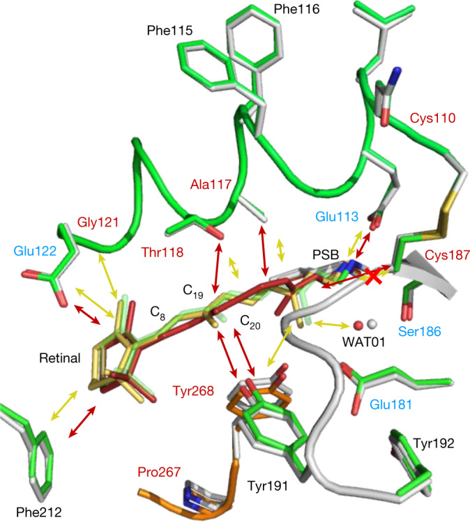
Fig. 4. Interactions between retinal and the binding-pocket residues are substantially reduced 1 ps after photoactivation.

Schematic of the interactions of retinal in the rhodopsin ligand-binding pocket before (red arrows) and after photoactivation in the picosecond range (yellow arrows; the red cross on the yellow arrow shows the bond disruption). A longer arrow represents a stronger interaction. The grey structure corresponds to the dark state (retinal in red) and the coloured structure corresponds to the 1-ps illuminated model (retinal in yellow). For comparison, the retinal model after 100 ps is shown in green. The residues labelled in red are GPCR-conserved and the blue residues are rhodopsin-conserved.
