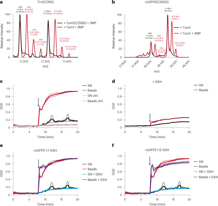
Fig. 5. Tum1 directly persulfidates other proteins.
a,b, Mass spectra of H. sapiens Trx1(C35S) (a) and roGFP2(C205S) (b) exposed to Tum1 and 3MP (red curve), or to an inactive Tum1 system (black curve). n = 1. c, Oxidation of roGFP2 (420 nM) by immobilized Tum1-SSH, in the absence of LMW compounds (beads; red line), or by the corresponding supernatant (SN; black line) of the reaction between immobilized Tum1 (2 µM) and 3MP (100 µM), that is, in the absence of Tum1-SSH. Control (ctrl) experiments were performed in absence of 3MP. n = 2 independent experiments. d, The same experiment as in c, but with the initial reaction between immobilized Tum1 (2 µM) and 3MP (100 µM) conducted in the presence of GSH (100 µM), thus diminishing formation of MPST-SSH. n = 2 independent experiments. e, Oxidation of roGFP2 (420 nM) by immobilized Tum1-SSH in the presence of GSH (beads + GSH; purple line), or by the corresponding supernatant in the presence of GSH (SN + GSH; blue line). Left panel: 420 nM GSH. Right panel: 2,100 nM GSH. The curves obtained in c (beads and SN in the absence of GSH; red and black lines, respectively) are included for direct comparison. n = 2 independent experiments.

