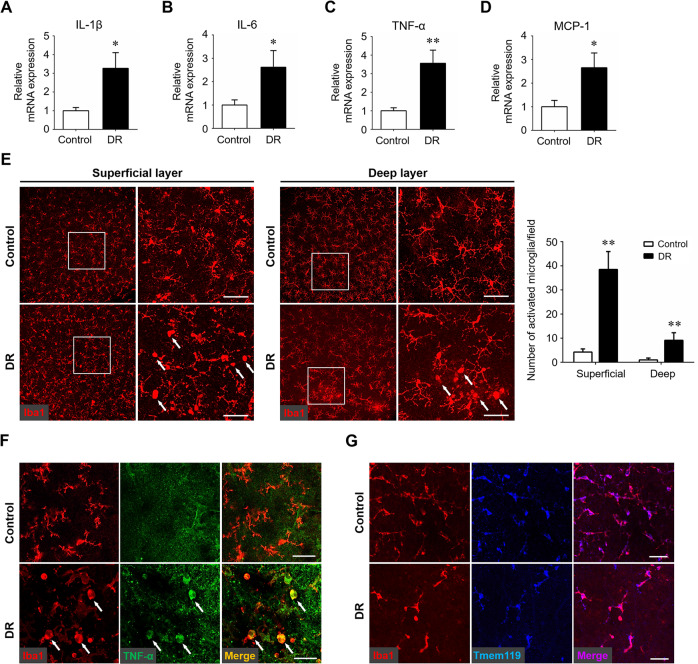
Fig. 1. Microglial activation and neuroinflammation cascade in the early stages of diabetic retinopathy.
Streptozotocin-induced diabetes model was established and retina samples were collected 12 weeks after diabetes induction. Age-matched non-diabetes mice were used as control. A–D qRT-PCR array was carried out for several inflammation-related genes, including IL-1β (A), IL-6 (B), TNF-α (C), and MCP-1 (D). Data were shown as mean ± S.D. The experiments were repeated 3 times for statistical analysis. E Representative images of iba-1 immunostaining on retinal flat-mounts. Arrows indicated activated microglia characterized by swollen cell bodies and short branched processes. Scale bar: 50 μm. Three images were obtained in different locations of the retina (central, mid-peripheral and peripheral areas) and 6 retinas from 6 mice in each group were used for analysis. F Immunostaining of iba1 and TNF-α on retinal flat-mounts. Arrows indicated cells co-stained with both iba1 and TNF-α. Scale bar: 50 μm. G Immunostaining of iba1 and Tmem119 on retinal flat-mounts. Scale bar: 50 μm. *P < 0.05, **P < 0.01.

