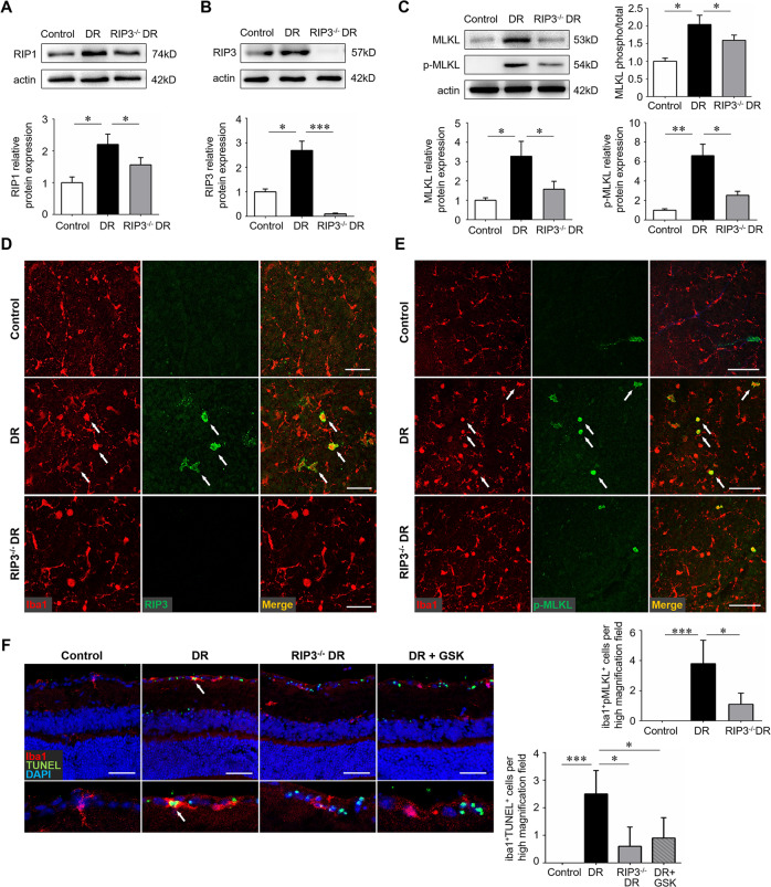
Fig. 3. RIP3 deficiency blocked the process of necroptosis in microglia.
Wild-type C57BL/6 J and RIP3−/− mice were used to create the streptozotocin-induced diabetes model and retinal samples were collected 12 weeks after diabetes induction. A, B The protein level of RIP1 and RIP3 was detected by western blotting. C The protein level of MLKL and p-MLKL were detected by western blotting. D Double immunofluorescence staining of iba1 and RIP3 on retinal flat-mounts. Arrows indicated cells that were both iba1 and RIP3 positive. Scale bar: 50 μm. E Double immunofluorescence staining of iba1 and p-MLKL on retinal flat-mounts. Arrows indicated cells that were both iba1 and p-MLKL positive. Scale bar: 100 μm. Three images were obtained in different locations of the retina (central, mid-peripheral and peripheral areas) and 6 retinas from 6 mice in each group were used for analysis. F Double immunofluorescence staining of iba1 and p-TUNEL assay on retinal cryosections. Arrows indicated iba1+ microglia that were undergoing cell death. Scale bar: 100 μm. Three horizontal sections were randomly chosen and 6 eyes from 6 mice in each group were analyzed. GSK, the specific RIP3 inhibitor GSK-872. Western blotting assays were repeated 3 times. Data were mean ± S.D. *P < 0.05, **P < 0.01, ***P < 0.001.

