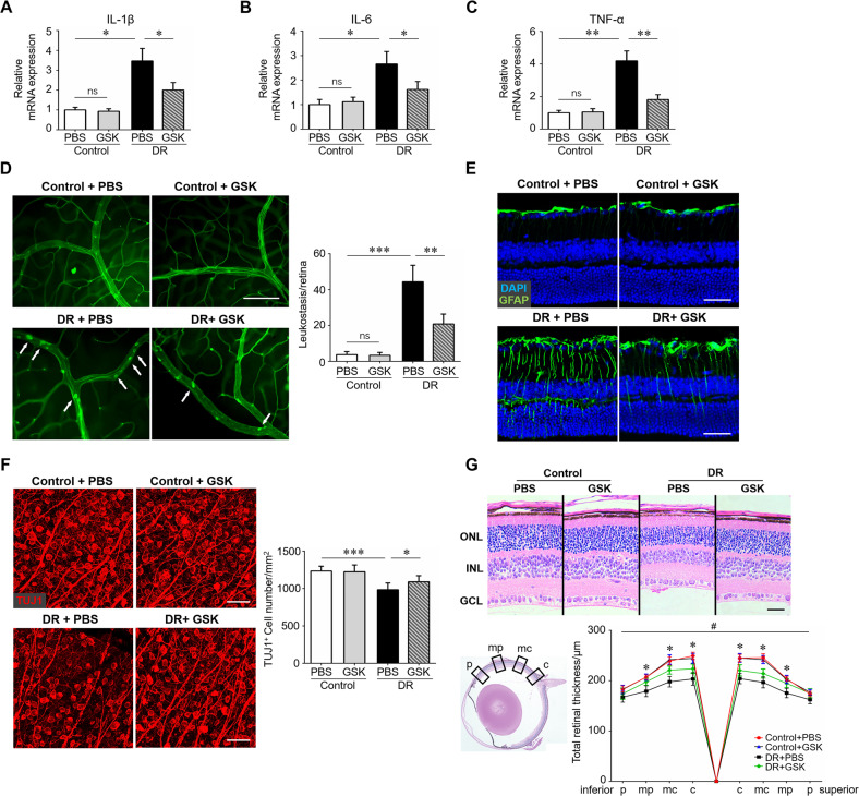
Fig. 4. RIP3 inhibition attenuated retinal neuroinflammation and neurodegeneration.
Streptozotocin-induced diabetes model was established. Age-matched non-diabetes mice were used as control. GSK-872 or vehicle were injected intravitreally on the first day of the 4th week and 8th week after STZ induction. Twelve weeks after diabetes induction, retina samples were collected and RNA was isolated using TRIzol Reagent. A–C qRT-PCR array was carried out for inflammation-related genes, including IL-1β, IL-6, and TNF-α. D Retinal leukostasis was performed using fluorescein-conjugated concanavalin A to label the adherent leukocytes. The number of adherent leukocytes were observed using a Leica microscope. Total adherent leukocytes per retina and six retinas from 6 mice in each group were used for analysis. Arrows indicated adherent leukocytes. Scale bar: 100 μm. E Immunostaining of GFAP, a marker for astrocytes and Müller cells, on retinal cryosections. Scale bar: 100 μm. F Immunostaining of TUJ1, a marker for neurons, on retinal flat-mounts. Scale bar: 50 μm. Three images were obtained in different locations of the retina (central, mid-peripheral and peripheral areas) and 6 retinas from 6 mice in each group were used for analysis. G H&E staining was carried out to evaluate retinal thickness in each group. Scale bar: 100 μm. Retinal thickness at different locations, including central (c), mid-central (mc), mid-peripheral (mp), and peripheral (p), were analyzed. Three horizontal sections were randomly chosen and 6 eyes from 6 mice in each group were analyzed. *DR + PBS vs DR + GSK, P < 0.05. GSK, the specific RIP3 inhibitor GSK-872. ONL outer nuclear layer, INL inner nuclear layer, GCL ganglion cell layer. Western blotting assays were repeated 3 times for statistical analysis. Data were shown as mean ± S.D. *P < 0.05, **P < 0.01. ***P < 0.001, ns no significance.

