. 2022 Sep 28;15(2):308–317. doi: 10.4055/cios22066

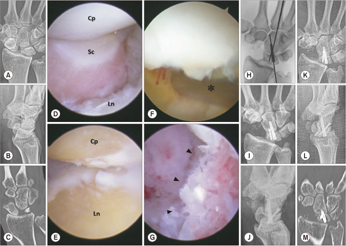
Fig. 1. Arthroscopic lunocapitate fusion in a patient with scapholunate advanced collapse (SLAC). (A-C) Preoperative posteroanterior and lateral radiographic views and a coronal computed tomographic scan of a 68-year-old man with stage II SLAC of the right wrist. (D) On the midcarpal ulnar portal (MCU), the scaphoid was dissociated from the scapholunate alignment (Cp: capitate, Sc: scaphoid, Ln: lunate). (E) The midcarpal radial portal (MCR) revealed the cancellous bone underneath the joint surface after curetting the cartilage and subchondral bone via the MCU as a working portal. (F, G) Using the MCR as a viewing portal and the MCU as a working portal, demineralized bone material (arrowheads) was inserted into the lunocapitate interval using a 3.5-mm burr sheath (asterisk) during fixation with temporary Kirschner wires. (H) On the intraoperative radiograph, temporary Kirshner wires were exchanged for headless compression screws. (I, J) Posteroanterior and lateral immediate postoperative radiographic views. (K-M) Posteroanterior and lateral radiographic views and a coronal computed tomographic scan at 10 weeks after surgery, showing that the lunocapitate interval achieved bony union.

Fig. 1