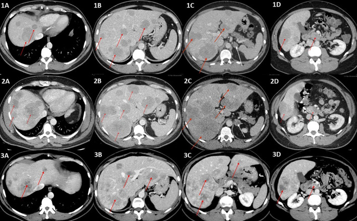
Figure 1.
(1A–1D) Contrast-enhanced CT scans of the abdomen (portal–venous phase) showing multiple metastases in the liver, ranging in size from a few millimeters to 40 mm (red arrows), incidentaloma of the left adrenal gland of benign phenotype 20 mm in diameter (white arrow), and primary tumor (red arrow) (1D). (2A–2D) Contrast-enhanced CT (portal–venous phase) scans revealed progression of the metastatic liver disease (red arrows) and a significant change in the left adrenal tumor density (47 HU in native phase) and size (24 × 18mm), arousing suspicion of adrenal metastasis (white arrow), which was not confirmed in final pathology reports. The primary tumor is indicated by a red arrow in (2D). (3A–3D) Post left-sided adrenalectomy contrast-enhanced CT scan showing stable metastatic liver disease, without further progression in terms of intensity, number, and size of metastases, and even with some of the hepatic metastases shrinking (red arrows). The primary tumor is indicated by a red arrow in (3D).

