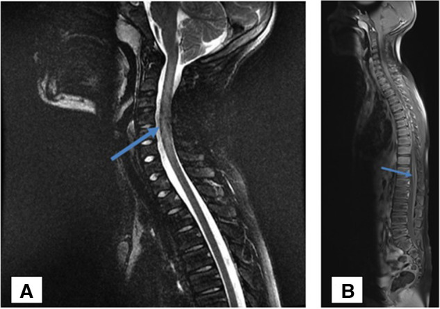
Figure 1.

MRI of the cervical spine showing extensive centromedullary T2 weighted image (WI) hypersignal (A) with normal T1 WI signal impairment between the C1–C7 metamers associated with mild swelling at the cervical spinal cord and leptomeningeal enhancement at the lumbosacral level and the cauda equine (B).
