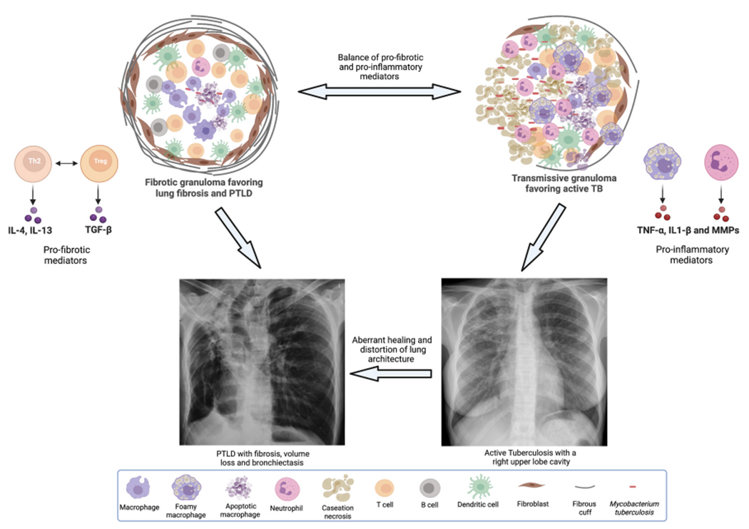
Fig. 1.

Depicts the characteristics of a fibrotic granuloma (left) and a transmissive granuloma (right). Fibrotic granulomas have an abundance of Th2 and Treg cells that drive TGF-β mediated collagen deposition, thick capsule consisting of myofibroblasts and an abnormal fibrous cuff. Transmissive granulomas are rich in classically activated and foamy macrophages as well as neutrophils. These cells produce inflammatory mediators such as tumor necrosis factor (TNF)-α and matrix metalloproteinases (MMPs) and depending on other host modulating factors, can result in the development of active cavitary TB disease. As the figure depicts, fibrotic granulomas favor the development of PTLD and transmissive granulomas favor the development of active TB, but granulomas can undergo changes driven by host factors and surrounding milieu such that they can morph into one or the other kind. Finally, the figure also depicts that active cavitary TB can lead to PTLD in the event of aberrant healing and distortion of lung architecture.
