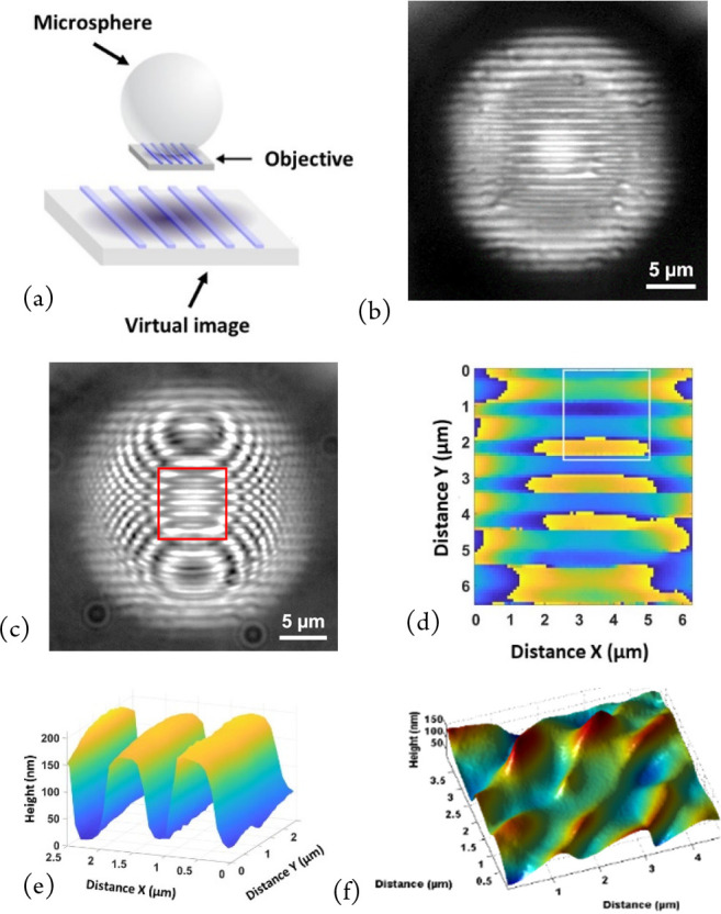
Figure 6.

Microsphere-assisted microscopy using 130 μm diameter glass microspheres: (a) Schematic drawing of virtual image formation. Reprinted with permission from Perrin, S.; Li, H.; Leong-Hoï, A.; Lecler, S.; Montgomery, P. C. Illumination conditions in microsphere-assisted microscopy. J. Microsc. (Oxford, U. K.)2019, 274, 1, 69–75. Copyright 2019 Wiley.20 (b) Focused on a 1.2 μm period grating through a 130 μm diameter microsphere, not visible directly with the ×20 objective, (c) fringes superimposed on an image of grating through a microsphere, (d) PSM measurement with phase discontinuities (central part from (b)), (e) topography measurement after removal of curvature due to spherical aberration (from (d)), and (f) nanoripples on stainless steel measured with a 25 μm diameter microsphere. Reprinted with permission from Leong-Hoï, A.; Hairaye, C.; Perrin, S.; Lecler, S.; Pfeiffer, P.; Montgomery, P. High resolution microsphere-assisted in-terference microscopy for 3D characterization of nanomaterials. Phys. Status Solidi A2017, 215, 1700858. Copyright 2017 Wiley.6
