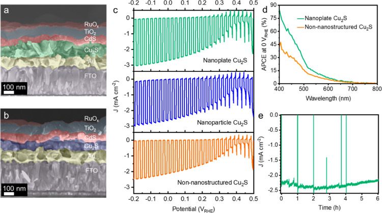
Figure 5.
(a) Cross-sectional SEM image of the Cu2S photocathode (FTO/Au/Cu2S/CdS/TiO2/RuOx) based on the nanoplate Cu2S thin film. (b) Cross-sectional SEM image of the Cu2S photocathode (FTO/Au/Cu2S/CdS/TiO2/RuOx) based on the nanoparticle Cu2S thin film. (c) J–E curves of Cu2S photocathodes based on different structures of Cu2S thin films under simulated on–off AM1.5 G illumination (100 mW cm–2). (d) Comparison of the APCEs of Cu2S photocathodes based on the nanoplate Cu2S thin film and the non-nanostructured Cu2S thin film. (e) Chronoamperometry of the Cu2S photocathode based on the nanoplate Cu2S thin film under constant bias at 0 VRHE under simulated AM1.5 G illumination (100 mW cm–2). All measurements were performed in a 1.0 M phosphate buffer solution (pH 7.0).

