
FIGURE 6.
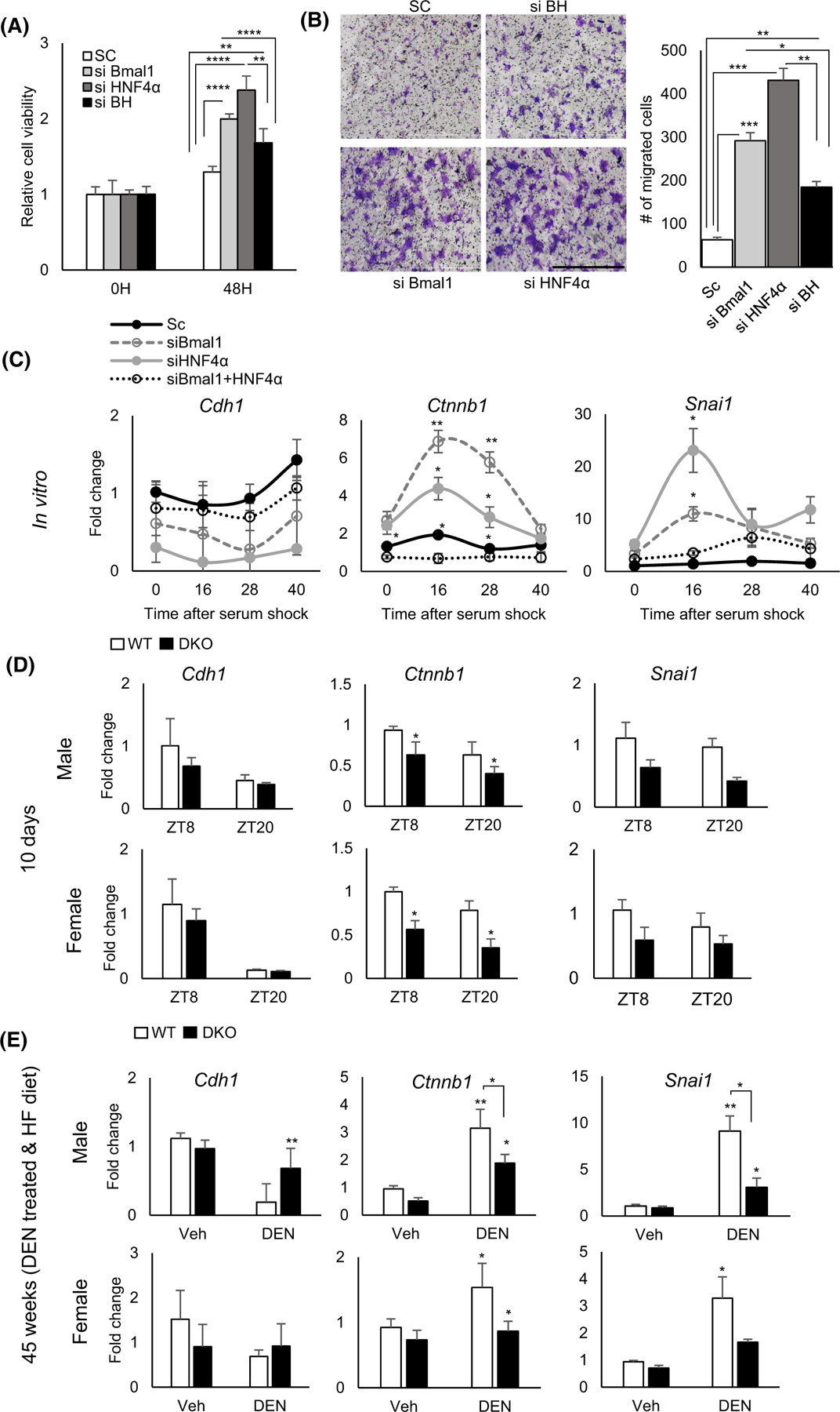
The Epithelial–Mesenchyme Transition (EMT) is impaired by dual loss of BMAL1 and HNF4α in the liver. (A) Fold change in proliferating Aml12 cells following HNF4α, Bmal1 or both Bmal1 and HNF4α knockdown using siRNA or scrambled control at 48 hr using MTT assay. (B) Migrated Aml12 cells following Hnf4α, Bmal1 or both Bmal1 and Hnf4α knockdown using siRNA or scrambled control. Quantification, left panel. (Scale bar = 1000 μm, N = 8). (C) qPCR reveals expression of EMT-related genes. in synchronized cells following Hnf4α, Bmal1 or both Bmal1 and Hnf4α using siRNA or scrambled control Two-way ANOVA, Sidak’s multiple comparisons test (N = 6–10): *p < .03; **p < .005; ***p < .0005, ****p < .0001 (D and E) Expression of EMT-related genes in BHLivDKO livers 10 days post tamoxifen injection (D) (Two-way ANOVA, Sidak’s multiple comparisons test [N = 6–8]: *p < .03) and in liver of BHLivDKO mice post VEH/DEN injection and 35 weeks of high fat diet feeding (E). Two-way ANOVA, Sidak’s multiple comparisons test (N = 6–8): *p < .03; **p < .005.