Figure 1:

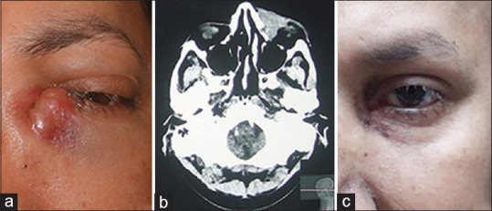
Clinical picture. (a) Acute swelling in the lacrimal area. (b) CT showing diffuse lesion in the lacrimal region extending to mid-orbit. (c) After receiving CHOP therapy and DCR operation. CT: Computed tomography, DCR: Dacryocystorhinostomy

Clinical picture. (a) Acute swelling in the lacrimal area. (b) CT showing diffuse lesion in the lacrimal region extending to mid-orbit. (c) After receiving CHOP therapy and DCR operation. CT: Computed tomography, DCR: Dacryocystorhinostomy