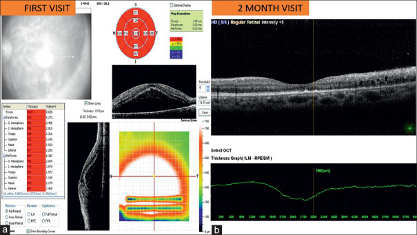
Figure 2:
Optical-coherence-tomography. (a) Left eye First-visit: neurosensory detachment. (b) Two months: Cystoid macular edema, subfoveal hyperreflectivity, and disruption of the external limiting membrane, the ellipsoid zone, and interdigitation zone in the foveal region. Features suggestive of acute macular neuroretinopathy

