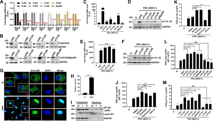
FIG 5.
Knockdown of TLR2, TLR3, TLR4, TLR5, and Myd88 enhances PRV replication. (A, B) Mouse peritoneal macrophages were transfected with control siRNA (siNC) and siRNAs targeting Tlr2, Tlr3, Tlr4, Tlr5, Tlr7, Tlr8, Tlr9, and Myd88 genes for 48 h or 72 h. The mRNA and protein levels of Tlr2, Tlr3, Tlr4, Tlr5, Tlr7, Tlr8, Tlr9, and Myd88 genes were detected with qPCR (A) and Western blotting (B). (C to F) Mouse peritoneal macrophages were transfected with control siRNA (siNC) and siRNAs targeting Tlr2, Tlr3, Tlr4, Tlr5, Tlr7, Tlr8, Tlr9, and Myd88 genes for 48 h and then mock infected or infected with PRV at MOI of 1 for another 24 h. The mRNA and protein levels of pro-IL-1β were detected with qPCR (C, E) and Western blotting (D, F). (G to I) Mouse peritoneal macrophages were infected with PRV for 24 h. (G) The subcellular locations of NF-κB (green) and the nuclear marker DAPI (blue) were visualized with confocal microscopy. (H) The percentage of the NF-κB localized in the cellular nucleus was calculated. The scale bars represent 5 μm. (I) The cell lysates were used for cytoplasmic and nuclear separation and analyzed by Western blotting. (J to M) Referring to the methods of panels C to F, PRV genomic DNA and the TCID50 were detected. Results shown are representative of three independent experiments (mean ± SD) or are representative of three independent experiments with similar results (one-way ANOVA) in panels C, E, and J to M. *, P < 0.05; **, 0.001 < P < 0.01; ***, P < 0.001; ns, no significance.

