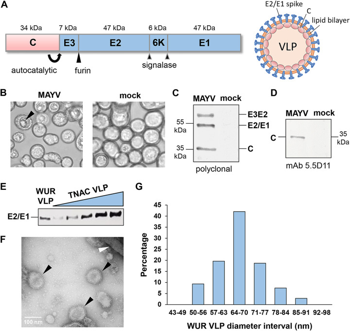
FIG 1.
MAYV VLP production using insect cells and recombinant baculoviruses. (A) Schematic overview of the MAYV structural cassette expressed in insect cells. The molecular weight of each protein is shown in kilodaltons. Autocatalytic, host furin, and host signalase cleavage sites are indicated. (B) BACe56/MAYV-infected Sf9 insect cells and mock-infected Sf9 insect cells at 4 days postinfection. Black arrow indicates dense nuclear body, which presumably consisted of accumulated MAYV core-like particles. (C and D) MAYV structural protein expression in Sf9 cells was analyzed at 4 days postinfection by western blotting using antiserum derived from a MAYV-infected mouse (C) or anti-CHIKV capsid antibody 5.5D11 (D). (E) Detection of MAYV structural proteins in purified WUR VLP fraction from Sf9 insect cells and dilution series of TNAC MAYV VLPs from HEK293 human cells. (F) Transmission electron microscopy photo of purified WUR MAYV VLPs. Black arrows indicate MAYV VLPs; white arrow indicates baculovirus. (G) Size distribution of WUR MAYV VLPs based on diameter measurements of 107 VLPs.

