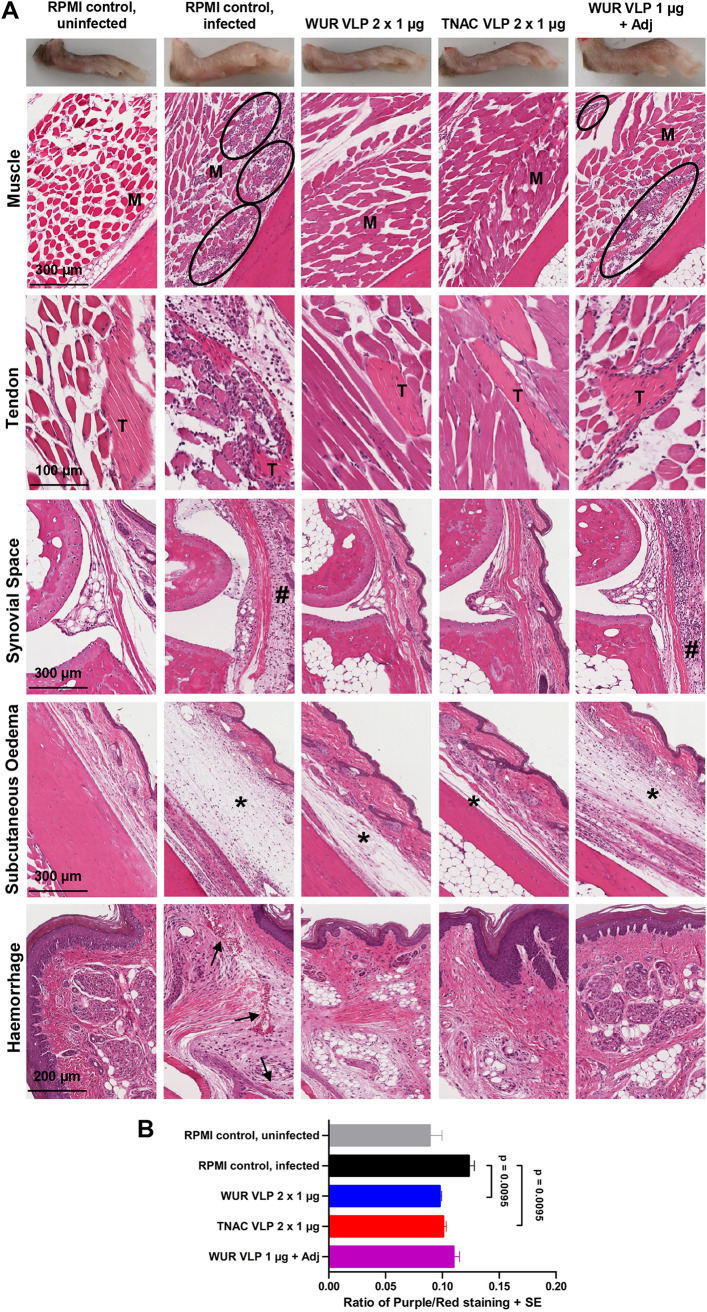
FIG 4.
Histopathology of MAYV VLP-vaccinated adult C57BL/6J mice after challenge with MAYV BeH407. (A) Photographs of mouse feet at 6 days postchallenge, illustrating swelling on day 6 in the RPMI-vaccinated, infected, and WUR VLP with adjuvant groups. H&E staining of tissues in foot sections from RPMI-vaccinated or VLP-vaccinated, uninfected, and MAYV-infected adult C57BL/6J mice is shown. Black ovals indicate some of the areas containing inflammatory infiltrates in the muscles (M). Inflammatory infiltrates near tendons (T) as well as inflammatory infiltrates near joint tissues (#) were also seen. Subcutaneous edema is shown with an asterisk. Hemorrhage is indicated by black arrows. These images are representative of a larger number analyzed. (B) Ratio of nuclear (purple) to nonnuclear (red) staining of H&E-stained foot sections (n = 2 to 3 mice, 4 to 6 feet per group, 3 sections per foot; values were averaged to produce one value for each foot). Statistical analysis used the Kolmogorov-Smirnov test. Multiple test correction was not applied.

