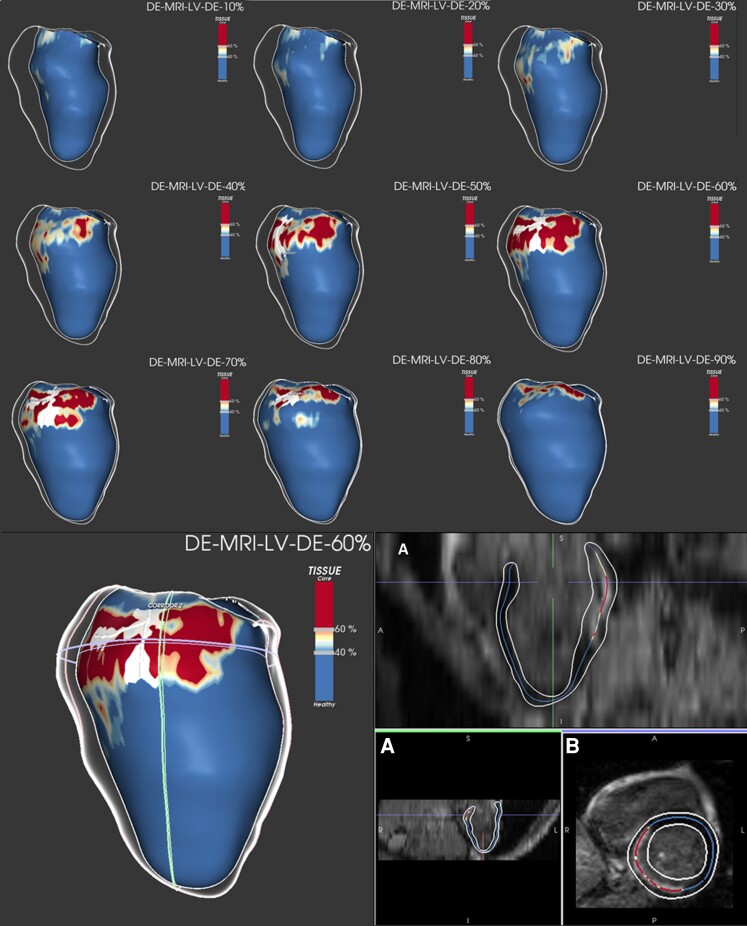
Figure 2.
LGE-CMR imaging post-processing with ADAS 3D software in a STEMI patient with BZ channel identification. Upper panel: LGE-CMR reconstruction of the LV showing an inferobasal scar of a STEMI patient (core in red, BZ in yellow, and healthy myocardium in blue). A white line is drawn over the surface, representing the BZ channel. The substrate progress is seen through different layers, from the endocardium (10–40%) to the epicardium (50–90%), with a defined BZ channel in different layers (50–80%). Lower left panel: LGE-CMR reconstruction of the same patient with the substrate seen from the epicardium (layer 60%). Lower right panel: LGE-CMR raw images in long (A) and short axis (B) used by the software to detect scar, core, BZ, and BZ channels (core in red, BZ in yellow, and healthy myocardium in blue). BZ, border zone; LGE-CMR, late gadolinium enhancement cardiac magnetic resonance; LV, left ventricle; STEMI, ST-segment elevation myocardial infarction;

