
Figure 3.
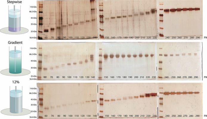
SDS-PAGE analysis of fractions collected from preparative gels. The experiments were carried out to optimize the gel concentration to achieve sufficient resolution for the separation of albumin from other proteins. Three different separating gel formats were tested; a stepwise gel composed of 1.5 cm 15%, 3 cm 12% and 1.5 cm 10% polyacrylamide gel layers and an 8% – 15% gradient gel and a 12% polyacrylamide gel. The total length of each separating gel was kept at 6 cm. Each separating gel was also topped with 1 cm, 4% stacking gel. The bottom of the preparative gel electrophoresis instrument was sealed with a dialysis membrane that had a molecular weight cut-off limit of 5 kDa. A total of 300 fractions, 0.5 mL of each, were collected. SDS-PAGE gels were silver stained and visualized with VersaDoc MP4000 imaging system.