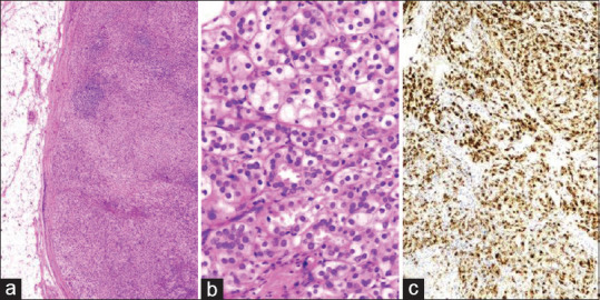
Figure 2.

(a) Photomicrograph of skin nodule showing tumor cells arranged in a nesting pattern (H and E, ×40), with occasional germinal center is identified. (b) High-power view shows the glandular architecture of the tumor with individual tumor cells having hyperchromatic nuclei and eosinophilic to clear cytoplasm (H and E, ×400). (c) The tumor cells showed diffuse cytoplasmic immunopositivity for PSA (IHC, ×100). H and E: Hematoxylin and eosin, IHC: Immunohistochemistry, PSA: Prostate-specific antigen
