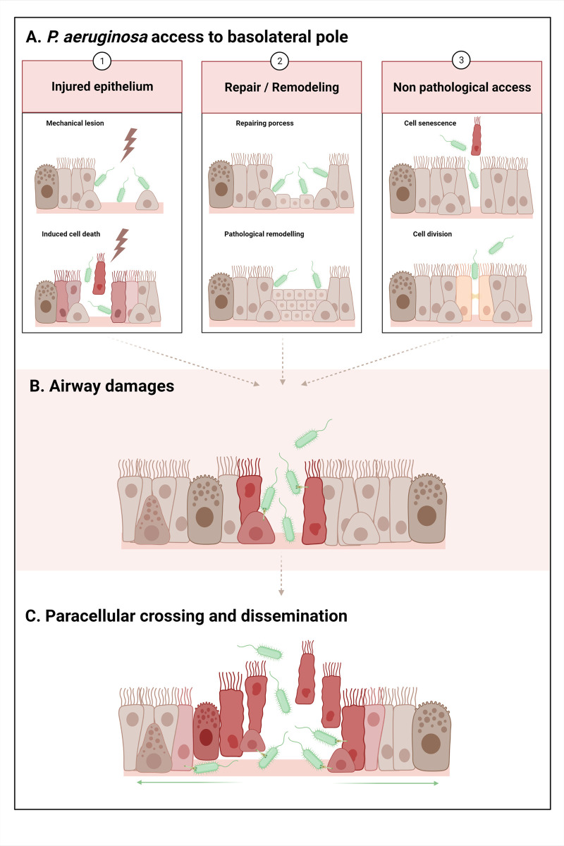
Fig 2. P. aeruginosa basolateral interaction and progression in the airway epithelium.
A. In the first step of infection, P. aeruginosa gains access to the basolateral part, exploiting various opportunities: 1. Injured epithelium: a. Mechanical lesion: removal of HAE cells and denudation of the basement membrane. Ex: endotracheal tube in VAP. b. Induced epithelial cell death: dying cells undergo retraction, tight junction disruption, and detachment from the adjacent cells and the basement membrane. The injury could be caused by pathogens (viruses, bacteria, etc.), chemical injury (pollutants, toxic compounds, etc.), or excessive inflammatory processes (excess of cytokines, proteases, oxidant stress, etc.). Ex: excess inflammation injuring HAE in CF and COPD. 2. Repair of the epithelium after injury/epithelium remodeling: a. After an injury, HAE undergoes a repair process: basal cell dedifferentiation, spread and migration, transitional squamous metaplasia or basal/mucous hyperplasia, and progressive differentiation. Cells display low differentiation levels, low polarity, and no functional tight junctions. Ex: repair in CF and COPD. b. Chronic and pathologically remodeled epithelium: squamous and goblet cells metaplasia, hyperplasia of surface goblet and basal cells. Cells display low differentiation levels, low polarity, and no functional tight junctions. Ex: remodeled epithelia in CF and COPD. 3. Nonpathological access in differentiated epithelium: transient disruption of tight junctions during: a. Extrusion of a senescent cell. b. Cell division. B. P. aeruginosa virulence factors induce airway damage, notably by T3SS toxin injection on the basolateral membranes of the cells. T3SS effectors (ExoS, ExoT, and ExoU) induce cell retraction and death, facilitating the subsequent access of bacteria to the adjacent or underlying cells and the basement membrane. C. Bacteria cross the epithelium by this paracellular route, gain access to the basal part, and progressively propagate radially through the epithelium to disseminate, using pili and flagella. Created with BioRender.com.

