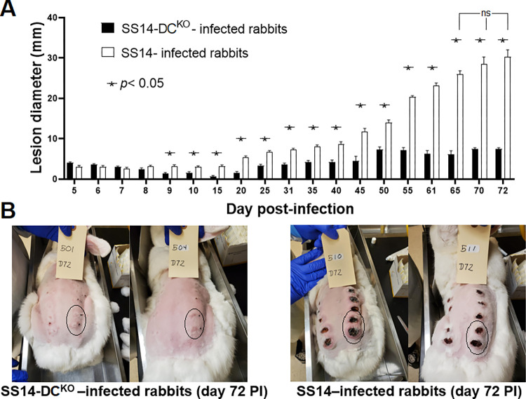
Fig 4. Lesion progression in infected animals.
(A) Measurements of lesion diameter (mm) over time in rabbits infected with the SS14 strain and the SS14-DCKO strain. Statistical analyses were performed using one-way ANOVA with the Dunnett correction of multiple comparisons or Student’s t-test, with significance set at p<0.05, indicated by an asterisk. The designation “ns” indicates lack of significance in lesion diameter of rabbits infected with the WT strain between day 65 and subsequent time points. (B) Pictures of the animals still in the study at day 72 post-infection showing large, ulcerated lesions in control rabbits (right panel), and significantly smaller (p<0.05) lesions in animals infected with the knockout strain (left panel). Circled areas represent lesions that were not yet biopsied at the time the picture was taken. Black ink dots near the lesion area mark the injection sites.

