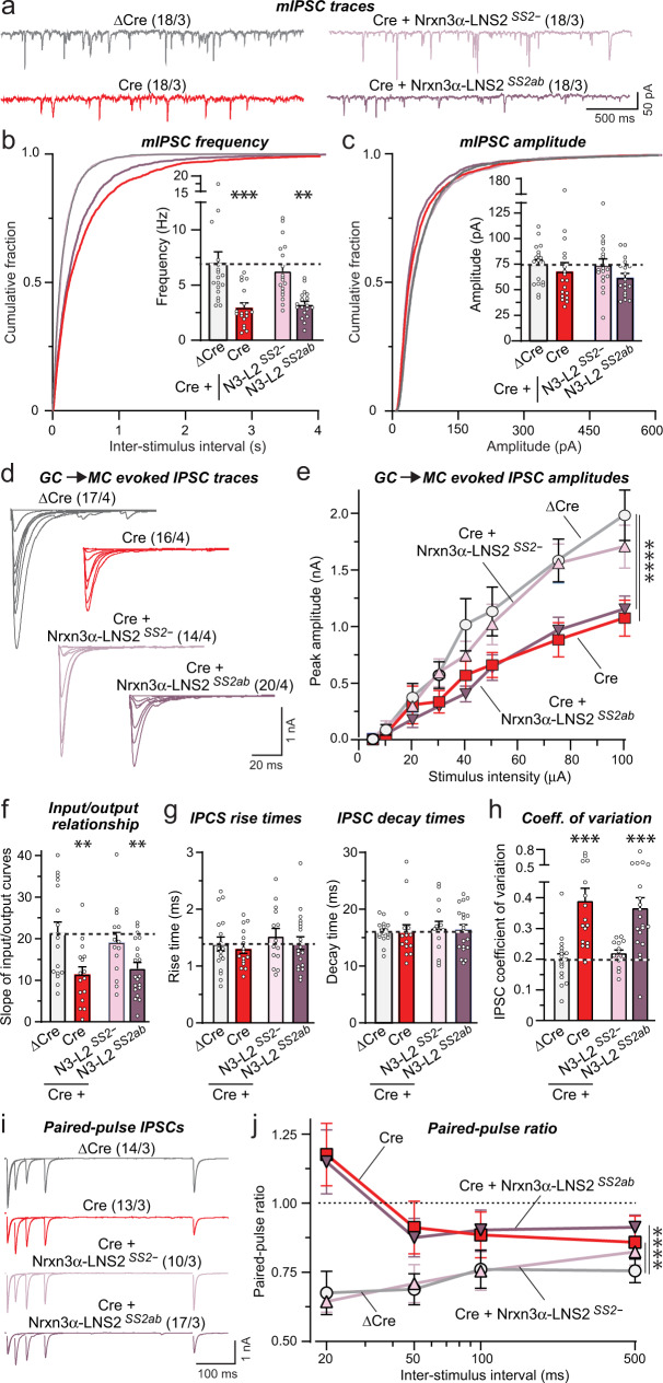
Fig. 4. Conditional in vivo deletion of Nrxn3 in the OB severely impairs GC→MC inhibitory synaptic transmission by lowering the release probability: Rescue by minimal Nrxn3-LNS2 constructs lacking an insert in SS2.
All experiments were performed by patch-clamp recordings from mitral cells in acute slices from Nrxn3 cKO mice whose OB was infected with AAVs (see Fig. 3a, S5c–e). a–c The Nrxn3 deletion decreases the mIPSC frequency; this decrease is rescued only by the minimal Nrxn3α-LNS2 construct lacking an insert in SS2 (a, representative mIPSC traces recorded in the presence of TTX; b, c cumulative probability of the mIPSC interevent intervals and amplitudes, insets: summary of the mIPSC frequency and amplitudes). d–f The Nrxn3 deletion decreases the evoked IPSC amplitude as documented by input/output curves. This decrease is rescued only by the minimal Nrxn3α-LNS2 construct without an insert in SS2 (d, representative IPSC traces; e, summary of input/output amplitudes; f, summary of the slope of input/output curves). g The Nrxn3 deletion and expression of rescue constructs have no effect on evoked IPSC kinetics (summary of the IPSC rise (left) and decay times (right)). h The Nrxn3 deletion increased the coefficient of variation of IPSCs, suggesting a decrease in release probability; this phenotype is rescued only by the minimal Nrxn3α-LNS2 construct without an insert in SS2. i, j The Nrxn3 deletion induces a large increase in the paired-pulse ratio; this phenotype is rescued by the minimal Nrxn3α-LNS2 construct without an insert in SS2 (i, representative traces; j, summary of the paired-pulse ratio). Numerical data are means ± SEM; n’s (cells/experiments) are indicated above the sample traces and apply to all graphs in an experimental series. Statistical analyzes were performed using two-way ANOVA in e and j and one-way ANOVA in b, c, and f–h with Dunnett’s and Tukey’s multiple comparison test respectively with regards to the ΔCre group, with *p < 0.05, **p < 0.01, ***p < 0.001, ***p < 0.001, and ****p < 0.0001. Source data and statistical results for all experiments are provided within the Source Data file.

