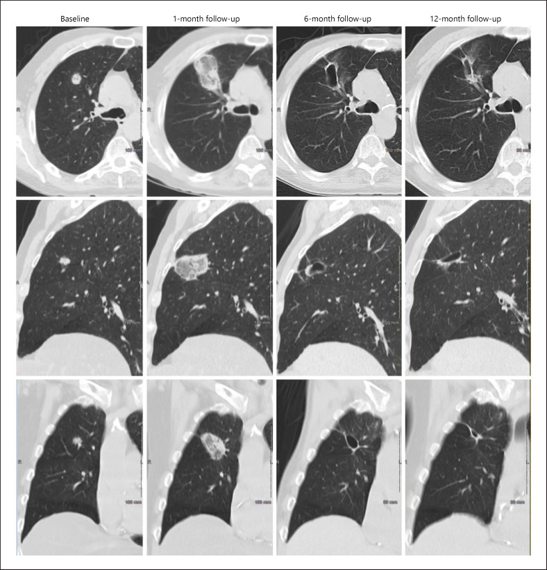
Fig. 3.
Serial CT images showing the evolution of an ablated target tumor over 12-month follow-up. The axial (top row), sagittal (middle row), and coronal (lower row) CT chest images from 1 representative patient from the study, at baseline (screening visit) (column 1) and at the 1-month (column 2), 6-month (column 3), and 12-month visit (column 4), are shown. Baseline images show a 14 × 13 mm target tumor in the upper lobe of the right lung. At month 1, the tumor is completely covered by the ablation zone and surrounded by a circumferential ablation margin (technique efficacy). At month 6 and 12, a small cavitary post-ablation zone is seen that contracts progressively over time, with no tumor recurrence evident.

