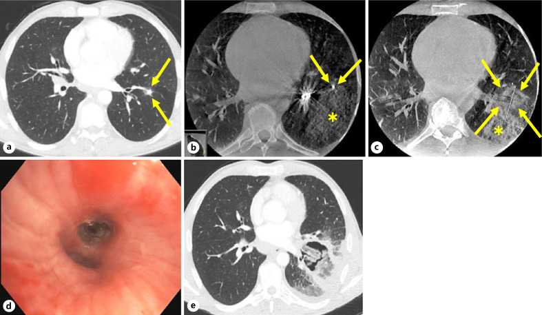
Fig. 4.
CT images of the patient who died unexpectedly 15 days post-ablation. The following are shown. a Baseline axial CT image taken at the screening visit, showing an 8 × 8 mm target tumor in the left lower lobe (arrows). b Axial CBCT image taken before ablation, showing the tip of the ablation probe (arrows) and limited parenchymal hemorrhage (asterisk). c Axial CBCT image 10 min after ablation, showing the tumor completely covered by the ablation zone (arrows) (estimation of ablation zone is difficult due to adjacent hemorrhage), with stable parenchymal hemorrhage (asterisk). d Bronchoscopic image taken after probe removal at the conclusion of the ablation procedure, showing intact bronchi and no blood. e Axial CT angiogram image taken on day 14 post-ablation at an outside hospital's emergency room showing a cavitary post-ablation tumor but no pseudoaneurysm, pulmonary embolism, or other vascular anomalies.

