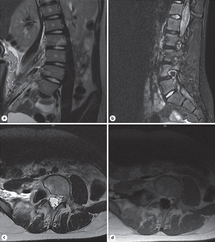
Fig. 2.
a Coronal T2: decompression of the right L4-L5 neural foramen, scalloping of the posterior aspect of the L4 vertebral body still present, no apparent residual tumor in the right paraspinal soft tissues and the psoas musculature is intact. b Sagittal STIR: slight reduction in edema in the posterior aspect of L4 vertebral body as well as the right posterior elements. c Axial T2: complete resection of the mass but widening the right L4-L5 neural foramen and scalloping the posterior aspect of the L4 vertebral body is still present, no residual mass seen in the right paraspinal soft tissues including the psoas musculature. d Post-contrast axial T1 shows no enhancement in the area where the lesion was indicating gross total resection.

