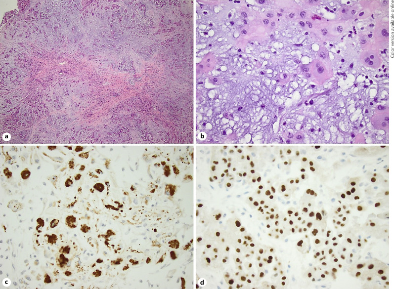
Fig. 3.
a Section of the tumor shows lobular pattern of short cords of eosinophilic epithelioid cells impeded in a myxoid stroma (H and E, ×40 magnification). b Section of the tumor highlights the physaliphorous cells along with the epithelioid tumor cells (H and E, ×400 magnification). c The neoplastic cells show immunoreactivity for pan-cytokeratin (×400 magnification). d The neoplastic cells show diffuse and strong nuclear staining for brachyury (×400 magnification).

