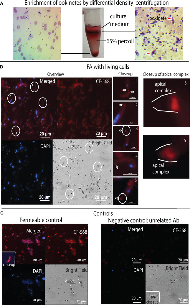
Figure 7.
Anti-α-tubulin Ab binds live P. falciparum ookinetes at their apical end. (A) Enriching ookinetes through differential density centrifugation using 65% Percoll. Arrows point ookinetes. (B) IFA assays localized α-tubulin-1 on living ookinetes. The co-localization of P. falciparum (nuclei, blue color) and α-tubulin-1 (red). A closeup of individual ookinetes shows Ab bound to the apical end of living P. falciparum ookinetes. A closeup of the apical complex shows anti-α-tubulin Ab bound to the apical polar ring of living ookinetes. (C) Ookinetes stained with CF568 dye-conjugated anti-V5 antibodies as a negative control showed no binding. Ookinetes fixed by methanol stained with anti-α-tubulin Ab, showing that Ab could stain α-tubulin-1 tubulin inside the permeable cells.

