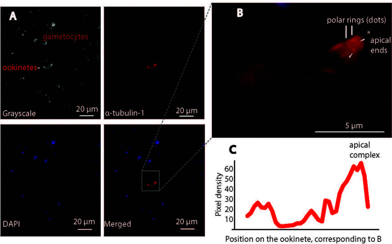
Figure 8.
Confocal immunofluorescence assays confirm P. falciparum α-tubulin-1 protein on the live ookinete surface. (A) α-tubulin-1 was detected on the live ookinete surface and not on the gametocyte surface. (B) Higher magnification showing the ookinete invasion apparatus of the ookinete (protrusion) and the strongest signals (red dots) evenly distributed at the apical polar rings (pointed by white arrows). * is the apical end. (C) Two rings at the apical region displayed the highest pixel density of red color.

