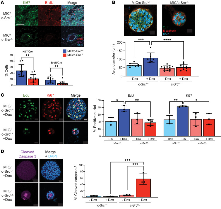
Figure 2. c-Src ablation impairs proliferation and increases apoptosis in an organoid model of early mammary tumor progression.
(A) Mammary glands from c-Src+/+ and c-SrcL/L MIC mice were immunostained with the indicated antibodies and DAPI. Representative images and quantification of Ki67+ and BrdU+ nuclei, normalized to Cre. n = 10 mice per group (minimum of 10,000 total nuclei per sample). Scale bar: 100 μm. **P < 0.01, by unpaired, 2-tailed Student’s t test. (B) Organotypic cultures were immunostained with the indicated antibodies and DAPI, and the organoid diameter was measured. Scale bar: 10 μm. ***P < 0.001 and ****P < 0.0001, by 1-way ANOVA with Tukey’s post hoc test. (C) Organoids were immunostained with the indicated antibodies (left panel), and staining was quantified and normalized to total nuclei (DAPI) (right panel). Scale bar: 10 μm. n = 3 independent mice per genotype (minimum of 20 total nuclei analyzed per mouse). *P < 0.05 and **P < 0.01, by 1-way ANOVA with Tukey’s post hoc test. (D) Organoids were immunostained to detect cleaved caspase 3 (left panel). Staining was quantified and normalized to the total number of cells, as determined by DAPI staining (right panel). n = 3 independent mice per genotype (minimum of 20 total nuclei analyzed per mouse). Scale bar: 10 μm. ***P < 0.001, by 1-way ANOVA with Tukey’s post hoc test. Dox, doxycycline.

