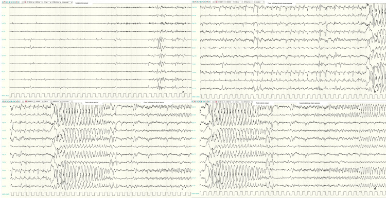
Figure 2. Ictal EEGs.
Ictal EEGs showed focal repetitive sharp waves on left central region corresponding to a focal clonic seizure (a); focal to bilateral repetitive sharp waves on centro-temporal regions (especially on the right) followed by generalized high amplitude spike and slow-wave discharge corresponding to a focal to bilateral tonic-clonic seizure (b-d).

