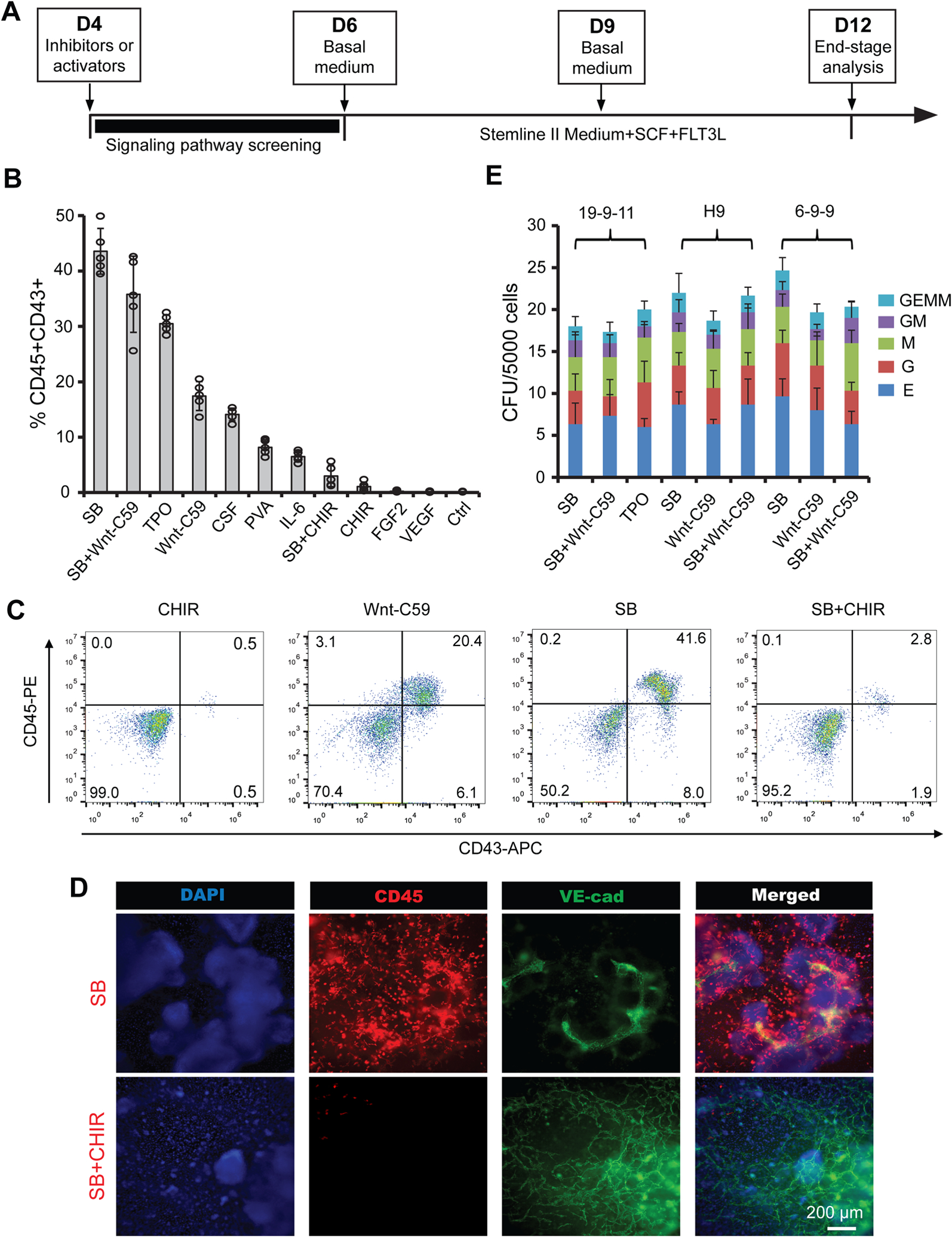
Figure 2.

Wnt and TGFβ inhibitors significantly induce hematopoiesis of hemogenic endothelium. (A) A schematic of the protocol used to differentiate CD34 + SOX17+ hPSC-derived hemogenic endothelium towards hematopoietic cells. (B–D) 19-9-11 iPSC-derived cultures differentiated as shown in (A) with indicated molecular signaling regulators were subjected to flow cytometry analysis for CD45/CD43 (B), and representative flow plots were shown in (C) and immunostaining images were shown in (D). CHIR, CHIR99021; SB, SB431542; Ctrl, control; PVA, polyvinyl alcohol; TPO, thrombopoietin; CSF, colony-stimulating factor 3. (E) Colony-forming unit (CFU) assay was performed on day 12 differentiated cultures with indicated treatment using 19-9-11, H9 and 6-9-9 hPSC lines, and scored by cellular morphology: erythroid (E), granulocyte (G), granulocyte/macrophage (GM), macrophage (M), and multilineage progenitor (GEMM). Data are represented as mean ± s. e.m of three to five independent replicates. Scale bars, 200 μm.
