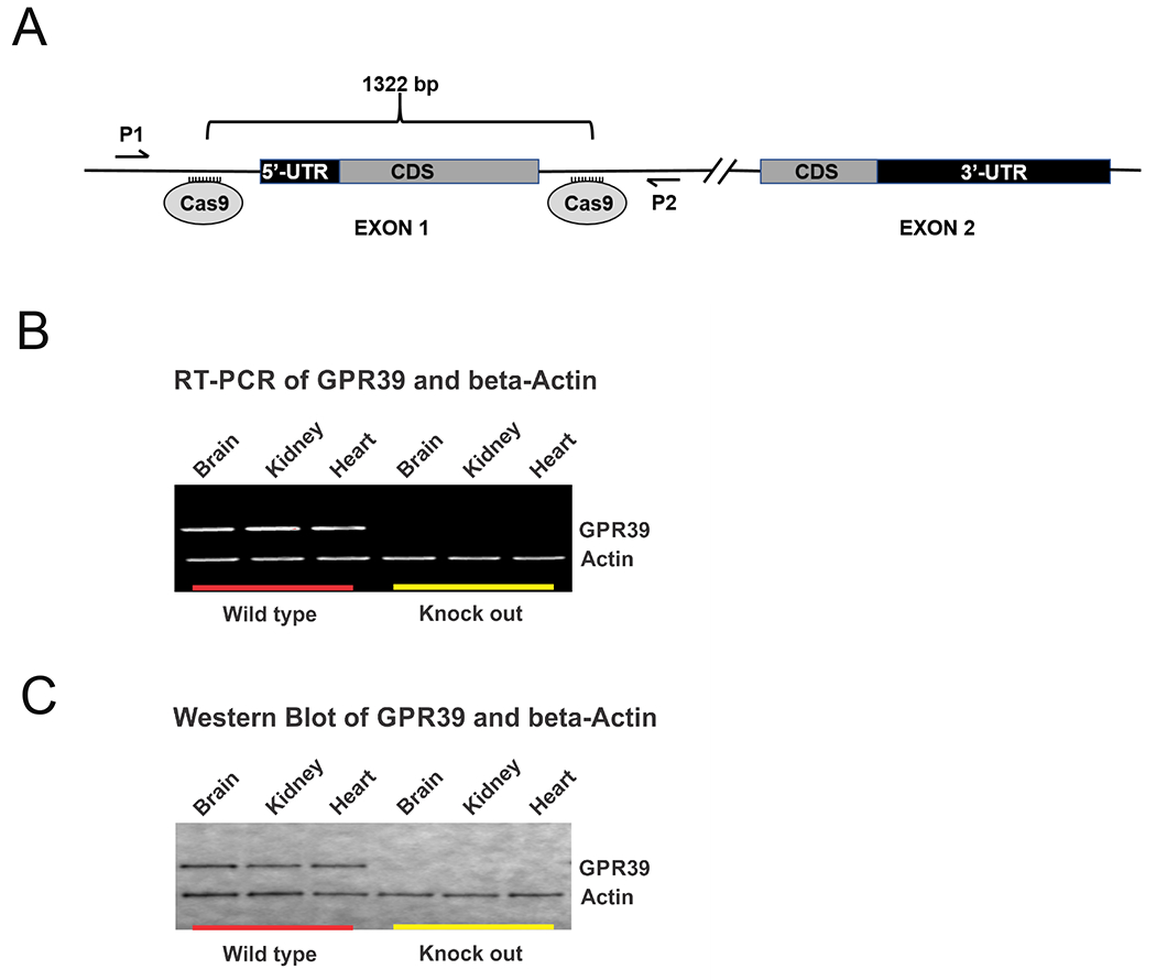
Figure 1. Creation and confirmation of mouse GPR39 knockout model.

A. Schematic of mouse GPR39 gene locus targeting strategy. Guide RNAs were designed to cleave at sites flanking exon 1 of Gpr39 resulting in a 1322 base pair deletion. Exon 2 was left intact. B. RT-PCR and C. Western blot of GPR39 (top row) and beta-Actin (bottom row) confirming presence of GPR39 mRNA (B) and protein (C) in brain, heart and kidneys from wild type mice (left) but absent in GPR39 knockout mice (right).
