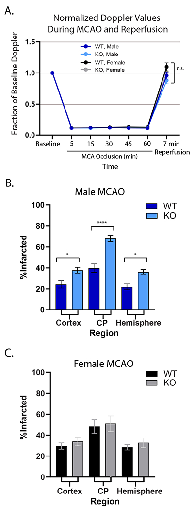
Figure 2. GPR39 deletion increases infarct size in male mice after MCAO.

A. Relative laser Doppler perfusion of MCA territory. There were no differences among groups. B. Infarct size was significantly larger in male GPR39 KO mice (n=14) compared to WT littermates (n=9) in cerebral cortex, caudate putamen (CP) and whole hemisphere. C. There was no difference in infarct size between WT (n=9) and GPR39 KO (n=12) in females. GPR39 KO vs. WT. n.s., not significant. *p<0.05, ** p<0.001. Cortex and CP are components of the hemisphere.
