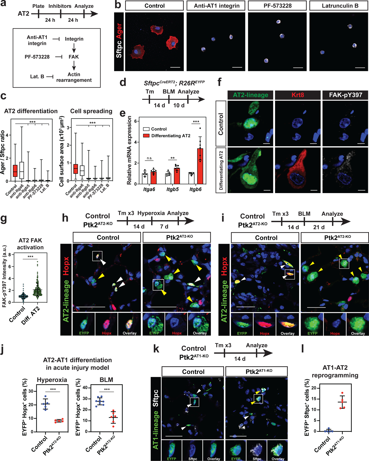
Figure 4. AT2-AT1 cell differentiation and AT1 cell fate maintenance require integrin signaling.

a-c. Integrin-FAK signaling is required for AT2 cell spreading and differentiation in vitro. Inhibitors were added 24 h after AT2 cell plating, and the cells were analyzed 24 h after the treatment (a). Anti-AT1 Integrin (Itgb6) antibody, FAK inhibitor PF 573228, or actin polymerization inhibitor Latrunculin B attenuated AT2 cell spreading and differentiation (b). The inhibitor-treated AT2 cells retained Sftpc+ Ager− AT2 cell state. Ager/Sftpc ratio and cell surface area were quantified for control (n = 135), anti-Itga6-treated (n = 69), anti-Itgb5-treated (n = 100), anti-Itgb6-treated (n = 126), PF 573228-treated (n = 123), or Latrunculin B-treated (n = 117) AT2 cells pooled from 3–6 independent experiments (c). d-g. Integrin-FAK signaling is activated during AT2-AT1 cell differentiation in vivo. Bleomycin (BLM) was intratracheally given to SftpcCreERT2; R26REYFP mice, and the mice were analyzed 10 days later (d). qPCR shows Itgb5, and Itgb6 are upregulated after acute lung injury, using sorted AT2 cells from PBS-treated control (n = 5 mice) and BLM-treated SftpcCreERT2; R26REYFP mice (n = 7 mice) from 3 independent experiments (e). FAK is activated (FAK-pY397+) in EYFP+ Krt8+ differentiating AT2 cells (f). FAK-pY397 intensity was quantified for n = 236 AT2 and n = 147 Krt8+ AT2 cells from n = 6 mice (g).
h-j. AT2-specific FAK knockout attenuates AT2-AT1 cell differentiation in vivo. FAK (Ptk2) deletion and lineage-tracing was performed on SftpcCreERT2; Ptk2flox/+; R26REYFP (control) and SftpcCreERT2; Ptk2flox/flox; R26REYFP (Ptk2AT2-KO) mice. The mice were analyzed 7 days after hyperoxia lung injury (h) or 21 days after BLM injury (i). AT2 cells differentiate into Hopx+ AT1 cells in control (h, i left), but less so in the Ptk2AT2-KO mice (h, i right) after injury. Quantification of AT2-AT1 cell differentiation for hyperoxia (n = 5 control and n = 5 Ptk2AT2-KO mice from 2 independent experiments) and BLM (n = 8 control and n = 6 Ptk2AT2-KO mice from 3 independent experiments) acute lung injury model (j).
k-l. AT1 cell-specific FAK deletion reprograms AT1 cells into AT2 cells. FAK deletion and lineage-tracing were performed on HopxCreERT2; R26REYFP (control) and HopxCreERT2; Ptk2flox/flox; R26REYFP (AT1Ptk2-KO) mice and analyzed 14 days later. AT1 lineage cells in AT1Ptk2-KO mice express AT2 cell marker Sftpc (k). Quantification of EYFP+ Sftpc+ reprogrammed AT2 cells from n = 5 mice (l).
*** P < 0.001 and ** P < 0.01 by one-way ANOVA (c) and two-tailed t-test (e, g, j). Each dot represents an individual mouse or cell, and error bars indicate mean with s.d. For Box and whisker plots, bars represent min and max values. Scale bars: b, h, i, k, 25 μm; f, 10 μm. See also Figure S5.
