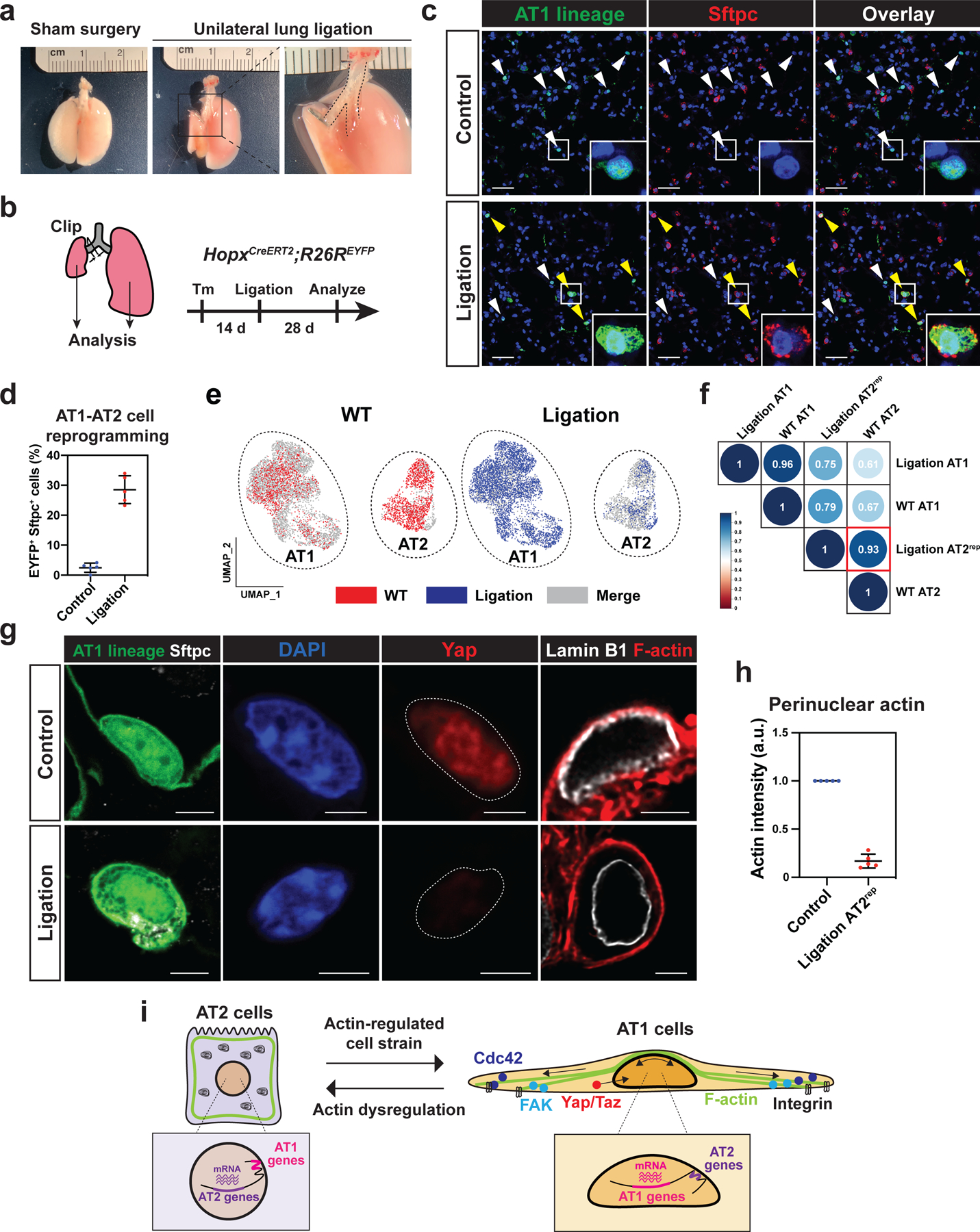
Figure 7. Alveolar cell fate is maintained by active breathing movements.

a-d. Unilateral lung ligation reprograms AT1 cells into AT2 cells. A gross morphological image of a mouse lung 28 days after sham surgery or unilateral lung ligation (a). The left main bronchus was ligated without affecting the left pulmonary artery or vein. Unilateral lung ligation and lineage-tracing were performed on HopxCreERT2; R26REYFP mice, and the mice were analyzed at 28 days later (b). Both the right (control) and left (ligated) lungs were analyzed. AT1 lineage cells express AT2 marker Sftpc in the ligated lung (c). White arrowheads indicate AT1 cells and yellow arrowheads indicate AT2rep cells. Quantification of EYFP+ Sftpc+ AT2rep cells from n = 5 mice from 3 independent experiments (d).
e-f. scRNA-seq analysis of wildtype alveolar epithelial cells and lineage-traced cells from HopxCreERT2; R26REYFP mice after unilateral lung ligation. AT2rep cells co-cluster with wildtype AT2 cells (e). Correlation analysis shows high similarity between AT2rep and wildtype AT2 cells (f).
g-h. YAP does not localize to the AT2rep nucleus and F-actin does not contact AT2rep nucleus. Representative images (g) and quantification (h) of perinuclear actin for control AT1 and AT2rep cells (n = 5 mice). Control lung and ligated left lung were adhered to the same glass slide. Actin intensity for AT2rep cells was standardized to that of control AT1 cells on the same glass slide.
i. Model of AT1 and AT2 cell regulation by biophysical forces.
Each dot represents an individual mouse, and error bars indicate mean with s.d. Scale bars: c, 25 μm; g, 2.5 μm. See also Figure S7.
