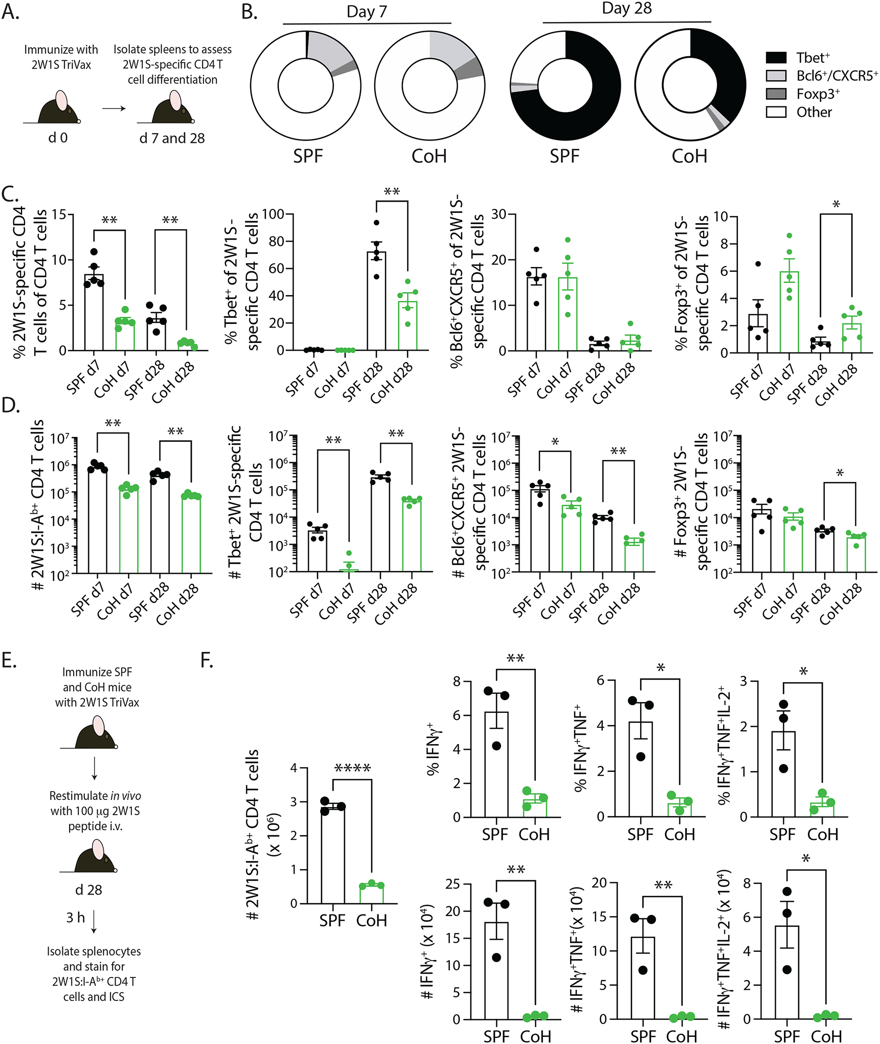
FIGURE 4. Reduced CD4 T cell differentiation and recall response in TriVax-immunized CoH mice.

A. Experimental design – SPF and CoH B6 mice were immunized with 2W1S-TriVax. On days 7 and 28 after immunization, spleens were isolated and processed for flow cytometry to determine the extent of 2W1S-specific CD4 T cell differentiation. B-D. The frequency and number of 2W1S-specific CD4 T cells that had differentiated into Th1 (Tbet+), Tfh (Bcl6+/CXCR5+), and Treg (Foxp3+) was determined. Representative flow plots are shown in Supplemental Figure 1. Data are representative of 2 technical replicates consisting of 5 mice/group/timepoint. * p ≤ 0.05, ** p ≤ 0.01. E. Experimental design – SPF and CoH B6 mice were immunized with 2W1S-TriVax. After 28 days, the mice were injected i.v. with 100 μg 2W1S56–68 peptide to restimulate the 2W1S-specific CD4 T cells to assess the ability to produce effector cytokines. Spleens were isolated 3 h later, and the cells were processed for flow cytometry. F. The frequency and number of IFNγ+, IFNγ+TNFα+, or IFNγ+TNFα+IL-2+ CD44+ 2W1S-specific CD4 T cells were determined by flow cytometry. Data are representative of 2 technical replicates, with 3 mice/group in each experiment. **** p ≤ 0.001, ** p ≤ 0.01, and * p ≤ 0.05.
