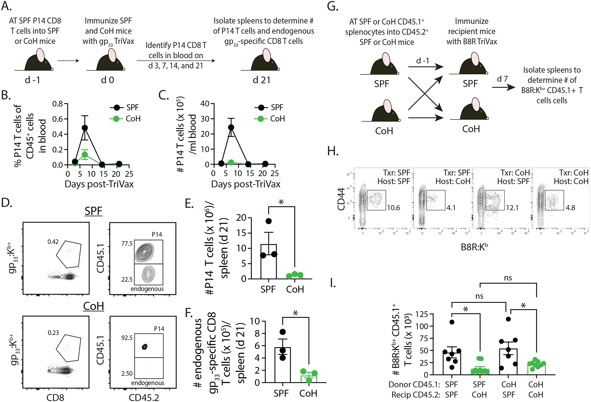
FIGURE 6. Differential expansion of SPF-derived P14 TCR-Tg CD8 T cells in TriVax-immunized SPF and CoH mice.

A. Experimental design – CD45.1+ P14 T cells were isolated from the spleens of P14 mice housed under SPF conditions, and then 5000 P14 CD8 T cells were adoptively transferred into SPF or CoH CD45.2+ B6 mice. The following day, the SPF and CoH recipient mice were immunized with gp33-TriVax. Blood was collected on days 3, 7, 14, and 21 post-immunization to determine the (B) frequency and (C) number of CD45.1+ P14 CD8 T cells among CD45.2+ cells. D-F. In addition, spleens were isolated on day 21 post-immunization to determine the number of (E) CD45.1+ P14 CD8 T cells and (F) endogenous CD45.2+ gp33-specific CD8 T cells. Representative flow plots are shown in panel D. * p ≤ 0.05. Data are representative of 2 technical replicates, with 3 mice/group in each experiment. G. Experimental design – 4 × 10⁶ bulk splenocytes from SPF or CoH CD45.1+ B6.SJL mice were adoptively transferred into SPF or CoH CD45.2+ B6 mice. The following day, the SPF and CoH recipient B6 mice were immunized with B8R-TriVax. H-I. Spleens were isolated from the recipient B6 mice 7 days after immunization to determine the number of CD45.1+ B8R-specific CD8 T cells. Representative flow plots are shown in panel H. * p ≤ 0.05. Data are combined from 2 technical replicates, with a total of 7–9 mice/group.
