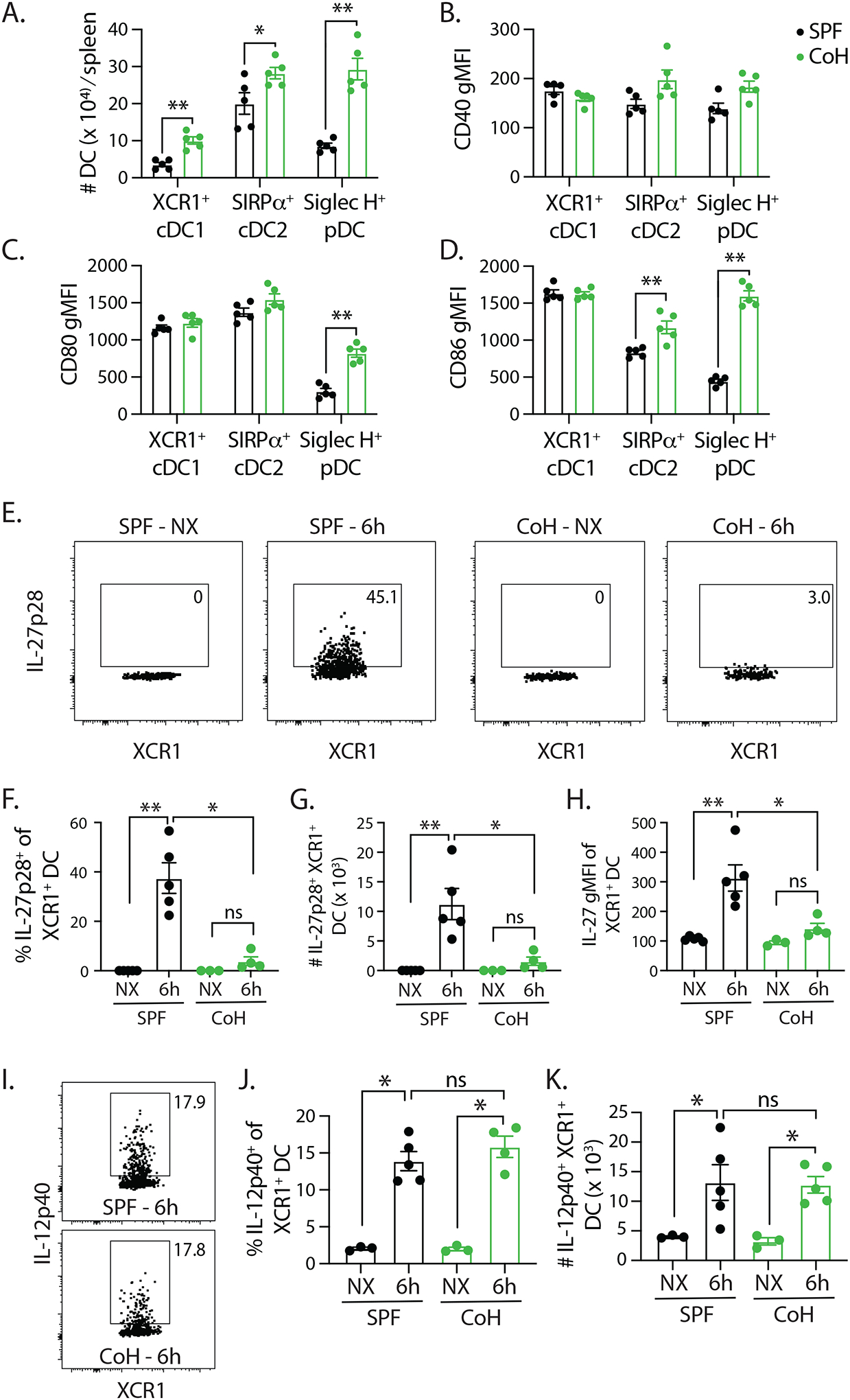
FIGURE 7. TriVax-immunized CoH mice have fewer IL-27p28-producing XCR1+ cDC1 cells.

A. Baseline numbers of XCR1+ cDC1, SIRPα+ cDC2, and Siglec H+ pDC in SPF and CoH mice. B-D. CD40, CD80, and CD86 expression was measured on each of these cell population, and the geometric mean fluorescence intensity (gMFI) is graphed. ** p ≤ 0.01, and * p ≤ 0.05. Data are representative of 2 technical replicates, with 3–5 mice/group in each experiment. SPF and CoH mice were immunized with B8R TriVax. Spleens were isolated 6 h later (as well as from unimmunized (NX) mice), and the expression of (E-H) IL-27p28 or (I-K) IL-12p40 by XCR1+ cDC1 cells was examined by flow cytometry. Representative flow plots show data from unimmunized and TriVax-immunized SPF and CoH mice. These data were used to determine the frequency and number of cytokine expressing XCR1+ cDC1 cells, as well as the IL-27p28 gMFI on the XCR1+ cDC1 cells.
