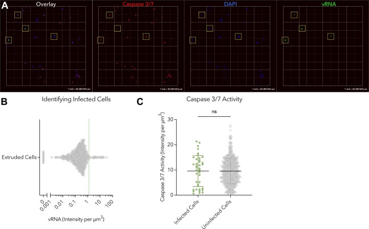
Extended Data Fig. 4. Extruded cells undergo anoikis by 48 hpi.
Cells that had fully extruded from infected colonoids in the presence of the fluorogenic caspase 3/7 substrate CellEvent were collected at 48 hpi, fixed, and examined by fluorescence microscopy at 20x magnification. (a) 3D renderings of imaged cells were visualized using Volocity image analysis software. Infected cells are surrounded by yellow boxes. (b) Individual cells were identified from 20 unique fields of view as in A using computational measurement tools in Volocity. Data were collected from N = 743 individual cells. Cells with pixel intensities greater than 1.5 per cubic micron in the vRNA channel were identified as infected; N = 39 infected cells, 5.2% of total. (c) The caspase 3/7 activity of cells defined in B were compared. Caspase activity in infected cells was comparable to that of uninfected cells; unpaired, two-sided t-test; data presented as mean ± SD; N = 39 infected cells; N = 704 uninfected cells.

