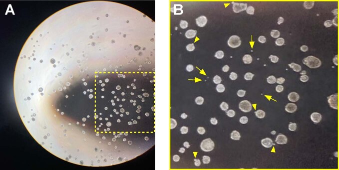
Extended Data Fig. 5. Examination of the Organoid fraction of suspension organoid cultures.
Following fractionation of organoid cultures, all fractions were visually examined on a tissue culture microscope to assess collection of target populations. (a) Visual examination of the Organoid fraction at 40x magnification confirms that organoids are collected with gravity pellet (1 x g). (b) Region highlighted in A. Single cells were also present at low abundance in the organoid fraction (arrows indicate examples), potentially a result of ongoing cell extrusion from organoids during fractionation (arrowheads).

