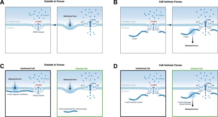
Extended Data Fig. 6. Potential mechanisms of mechanosensitive ion channel activation in infected cells.
We provide two hypotheses for potential means of activation of mechanosensitive ion channels in infected cells that may drive infected cell extrusion. Mechanosensitive ion channels such as Piezo1 can be activated by (a) external compression forces that induce membrane deformation, as well as (b) cell-intrinsic mechanical forces transmitted through tethered cytoskeletal filaments. (c, d) We present conceptual models for how viral infection may induce channel activation via both outside-in or cell-intrinsic forces. (c) In uninfected cells, cytoskeletal filaments provide structural support beneath the plasma membrane, reducing membrane deformation from cell-extrinsic forces. In infected cells wherein actin is rearranged, this structural support may be missing, resulting in ‘softer’ membranes more easily deformed by external compression forces. (d) In the force-through-filament model, cell-intrinsic mechanical forces are transmitted to Piezo channels via tethering to cytoskeletal filaments. In infected cells, cytoskeletal rearrangements may transmit forces directly to tethered Piezo channels, inducing activation. Adapted from ‘PIEZO Channels: How Do They Allow Mechanosensation?’, by BioRender.com (2022). Retrieved from https://app.biorender.com/biorender-templates.

