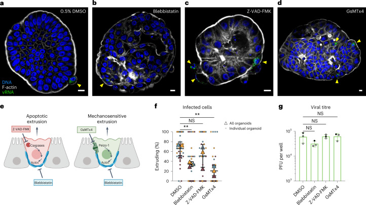
Fig. 4. Mechanosensitive signalling promotes infected cell extrusion.
EV-A71-infected organoids were exposed to compounds capable of inhibiting cellular factors implicated in different mechanisms of extrusion. a–d, Infected organoids were exposed to 0.5% DMSO vehicle control (a), 50 μM para-nitro-blebbistatin (b), 100 μM Z-VAD-FMK (c) or 20 μM GsMTx4 (d). Infected cells in organoids were visually inspected by confocal microscopy. Yellow arrowheads indicate infected cells in representative organoids. Scale bars, 10 μm. e, Blebbistatin inhibits both apoptotic and mechanosensitive extrusions, Z-VAD-FMK inhibits only apoptotic extrusion and GsMTx4 inhibits only mechanosensitive extrusion. f, The percentage of infected cells undergoing extrusion after 7 h of infection was enumerated. Each colour shows an independent experiment. Overall proportion of infected cells extruding per experiment shown as triangles, with measurements for each organoid shown as small circles. **P < 0.01; NS, not significant; repeated measures one-way ANOVA with Dunnett’s multiple comparisons test, N = 3. g, Viral titres quantified at 7 h post infection from infected suspension organoid cultures show no significant effects of drug treatments on virus yield. Repeated measures one-way ANOVA with Dunnett’s multiple comparisons test, N = 3 independent infections. In f and g, horizontal and error bars represent mean ± s.d.

