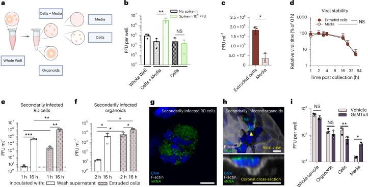
Fig. 5. Extruded cells can infect cultured cells and organoids.
a, EV-A71-infected colonoid cultures were collected at 8 h post infection, and components were collected by differential sedimentation. b, To assess effectiveness of washing extruded cells, 106 PFU exogenous free virus was spiked-in to Cells + Media samples. Fractions including Whole Well (blank bars), Cells + Media (small dotted bars) and Cells (large-dotted bars) were subjected to freeze–thaw and plaque assay. Repeated-measures one-way ANOVA with the Holm–Šídák multiple comparisons test. NS, not significant. c, Distribution of virus in Cells and in Media. Ratio paired, two-tailed t-test. d, Stability of virus from Cells and Media fractions. Collected fractions were incubated at 37 °C for the times indicated, subjected to repetitive freeze–thaw and subsequent plaque assay. Amounts of virus are normalized to values from c before incubation. Two-way ANOVA with Geisser–Greenhouse correction. e, Intact extruded Cells fractions and free virus from the final wash of Cells were used to infect RD cell monolayers. Viral titres in the infected RD cells were measured 1 h and 16 h after initiating secondary infection. Ratio paired (cells–cells; wash–wash) and unpaired (wash–cells), two-tailed t-tests. Dashed line indicates limit of detection. f, Cells fractions and free virus from the final wash of Cells were used to infect new colonoids. After 2 h and 16 h, the amount of virus in Whole Well fractions was determined by plaque assay. Statistic testing as described in e. g,h, Confocal microscopy of secondarily infected RD cells (g) and organoids inoculated with Cells after 16 h (h). Scale bars, 10 μm. h, Secondarily infected organoid with several infected cells. Bottom: orthogonal cross-section through the secondarily infected, extruding cell indicated by yellow arrowhead. i, EV-A71-infected colonoid cultures were treated with GsMTx4 or vehicle, and fractions were collected after 8 h. While GsMTx4 treatment reduced the amount of infectious virus in extruded cells, the amount of infectious free virus in the media increased. Multiple paired, two-tailed t-tests with the Holm–Šídák correction for multiple comparisons. In b–f and i, one representative experiment is shown with independent infections performed in triplicate; *P < 0.05, **P < 0.01, ***P < 0.001; data represented are mean ± s.d.

