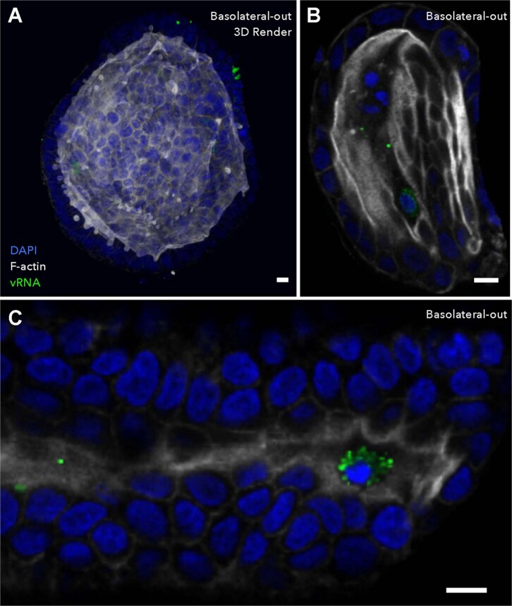
Extended Data Fig. 2. Apical extrusion of EV-A71 infected cells occurs regardless of organoid polarity.
Differentiated colonoids with basolateral-out organoid polarity were infected with EV-A71 and visualized by immunofluorescence at 8 hours post-infection. (a) Strong F-actin staining of the apical microvillus brush border was observed on the interior lumen of infected colonoids. Virus infected cells can be observed both within the epithelial layer, as well as within the apical lumen. (b, c) EV-A71 infected cells were apically extruded into the interior lumen of basolateral-out colonoids. Scale bars equal 10 μm.

