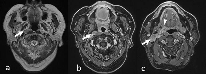
Fig. 19.
Incidentally detected mesopharyngeal carcinoma (p16 negative) with lymph node metastases in a patient with lung adenocarcinoma. T2-weighted image a shows right retropharyngeal lymph node metastasis as a hyperintense nodule in the retropharyngeal area (arrow). Contrast-enhanced 3D T1-weighted images with fat suppression of axial reconstruction (b and c) show an enlarged right retropharyngeal lymph node (b, arrow), right superior internal jugular nodule (c, arrow), and right mesopharyngeal tumor (c, arrowhead)

