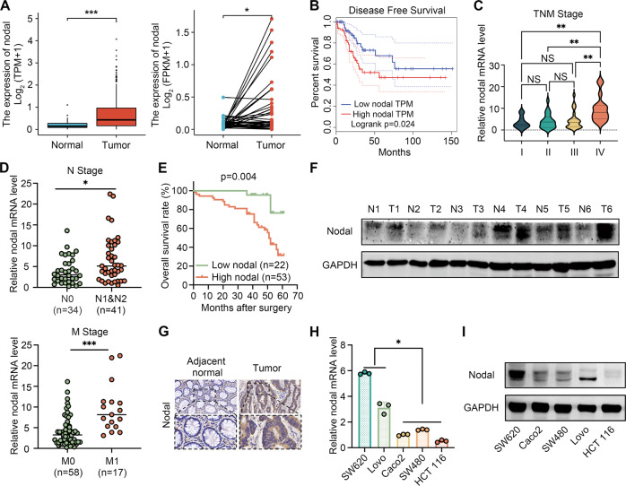
Fig. 1. Nodal expression is elevated in CRC, which is associated with the metastasis and poor clinical outcomes of CRC.
A Expression profile of Nodal in CRC tissues in TCGA cohort. B Increased Nodal expression was associated with poor disease-free survival in CRC tissues in TCGA cohort. C, D Increased Nodal expression was associated with an advanced TNM stage, lymph node metastasis and distant metastasis in 75 CRC tissues obtained from patients. E Kaplan–Meier analysis (log-rank test) for the overall survival of 75 patients with CRC in the high- and low-Nodal-expression groups stratified based on the median Nodal expression. F Western blotting was performed to evaluate the protein expression of Nodal in six pairs of CRC tissues. G Representative images of immunohistochemical (IHC) staining for Nodal in CRC tissues and their adjacent normal tissues (magnification, 200×). H, I qRT-PCR and western blotting were performed to examine the mRNA and protein expression of Nodal, respectively, in several CRC cell lines. (metastatic cell lines: SW620 and Lovo; non-metastatic cell lines: Caco2, SW480 and HCT116). Data are expressed as mean ± SD of three independent experiments (∗P < 0.05, ∗∗P < 0.01, and ∗∗∗P < 0.001).

