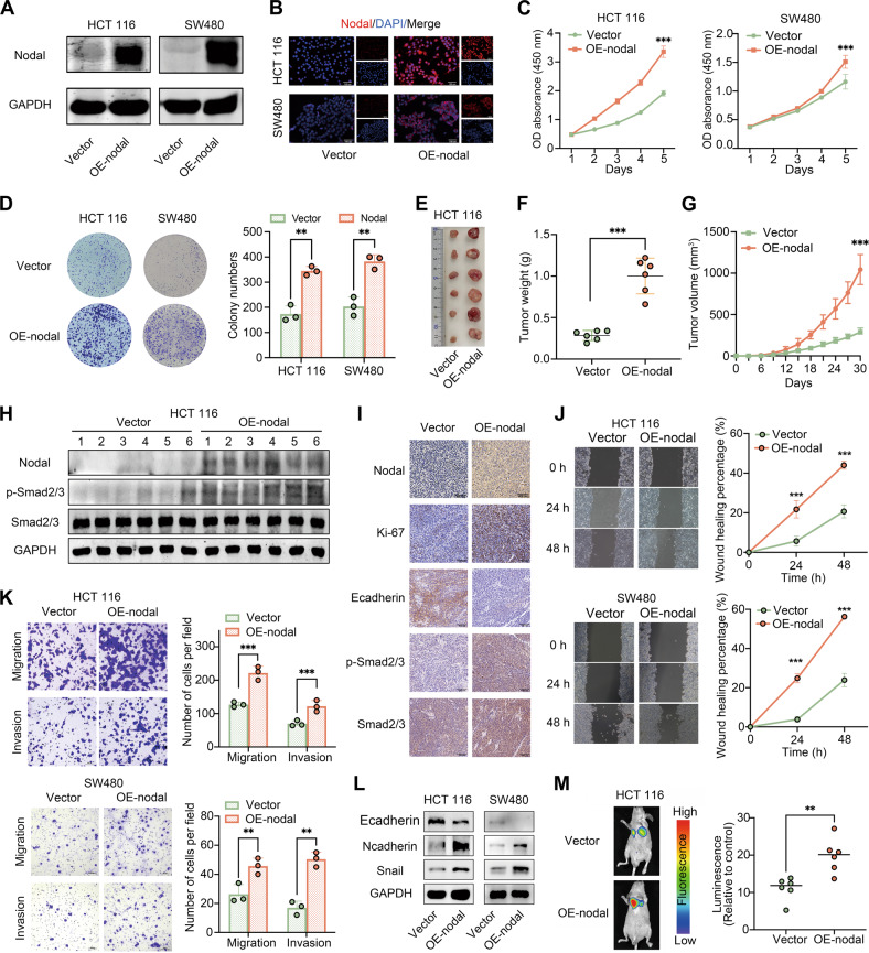
Fig. 2. Overexpression of Nodal promoted the proliferation, migration and invasion abilities of CRC cells in vitro and in vivo.
A Western blotting was performed to examine the protein expression of Nodal in Nodal-overexpressing HCT116 cells and SW480 cells (vector group: cells transfected with an empty vector; oe-Nodal group: cells transfected with the Nodal lentiviral overexpression vector). B Representative images of Nodal expression detected in Nodal-overexpressing HCT116 cells and SW480 cells via IF staining. C, D CCK-8 and colony formation assays validated the increased proliferative ability of HCT116 and SW480 cells after Nodal overexpression. E Representative images for the xenograft tumours morphology. F, G The volume and weight of tumours were evaluated in two groups. H Western blotting was performed to examine the expression of Nodal, p-Smad2/3 and Smad2/3 in each xenograft tumor tissue. I The Nodal, Ki-67, E-cadherin, p-Smad2/3, and Smad2/3 expressions in xenograft tumour tissues were detected via IHC staining. J Wound healing assay revealed that Nodal overexpression increased the migration ability of HCT116 and SW480 cells. K Transwell assay validated the increased migration and invasion ability of HCT116 and SW480 cells after Nodal overexpression. L Western blotting was performed to examine the E-cadherin, N-cadherin and Snail expressions of HCT116 and SW480 cells in the vector/oe-Nodal group. M Representative fluorescence images of lung metastasis in mice and the relative luminescence intensity. (∗∗P < 0.01 and ∗∗∗P < 0.001).

