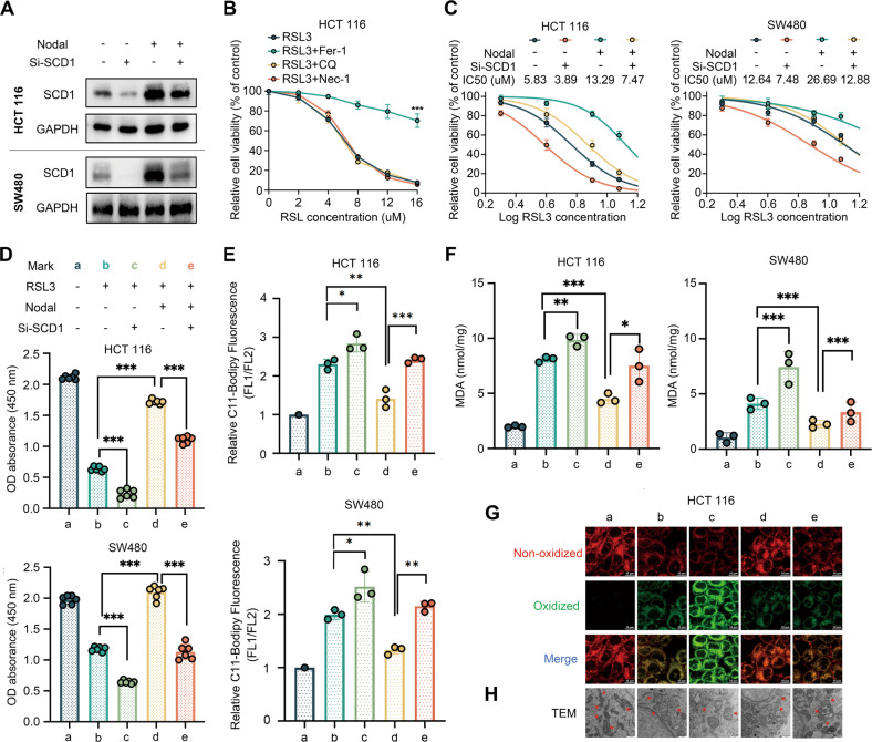
Fig. 5. Nodal regulated the sensitivity to RSL3-induced ferroptosis via SCD1.
A Western blotting of SCD1 in HCT116 and SW480 cells in the vector and oe-Nodal groups with or without si-SCD1 transfection. B Relative cell viability of HCT116 cells treated with different concentrations of RSL3 (a ferroptosis inducer) for 48 h with or without several cell death inhibitors. C Relative cell viability of HCT116 and SW480 cells in the vector and oe-Nodal groups with or without si-SCD1 transfection. The cells were treated with different concentrations of RSL3 for 48 h. D Cell viability was measured via CCK8 assay in HCT116 and SW480 cells in the vector and oe-Nodal groups with or without si-SCD1 transfection. HCT 116 cells and SW480 cells were treated with 8 μM and 12 μM RSL3 for 48 h, respectively. E BODIPY™ C11 staining analysis of lipid peroxidation via flow cytometry in Nodal-overexpressing HCT116 and SW480 cells with or without si-SCD1 transfection. F MDA levels in HCT116 and SW480 cells in the vector and oe-Nodal groups with or without si-SCD1 transfection. G Representative confocal laser scanning microscopic images of lipid peroxidation in HCT116 cells (green: oxidised lipids; red: non-oxidised lipids). H Representative transmission electron microscopic images of morphological changes in mitochondria of HCT116 cells (red arrows: mitochondria). (∗P < 0.05, ∗∗P < 0.01 and ∗∗∗P < 0.001).

