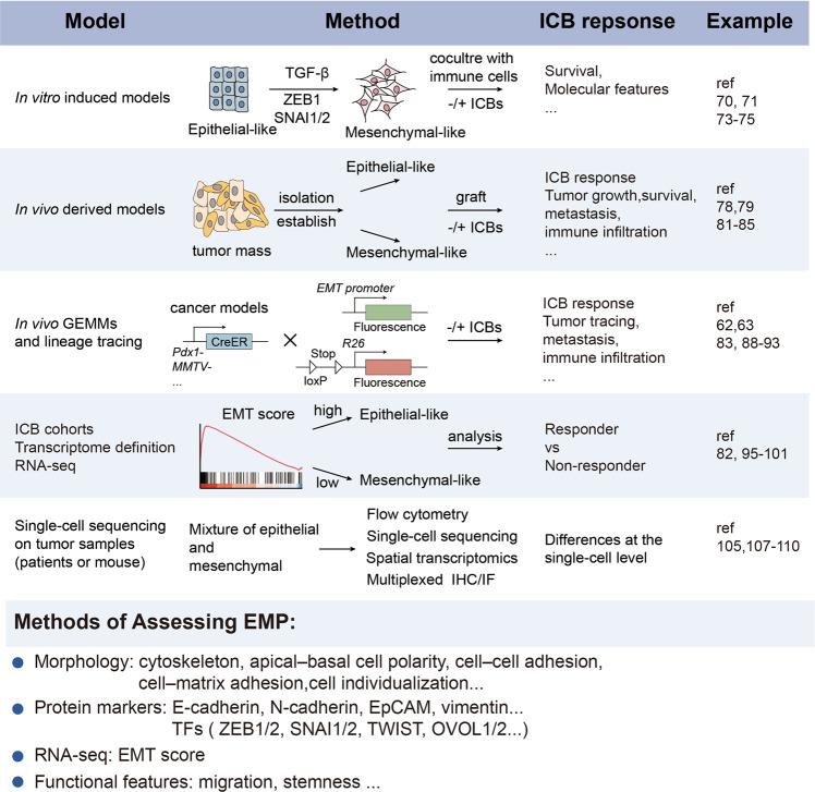
Fig. 2.
Advanced methods to study the link between EMP and the ICB response. The schematic diagram illustrates the advanced methods and models used to clarify the relationship between the ICB response and diverse EMP states in tumors. (1) In vitro induced models: 2 types of cancer cells (epithelial-like or mesenchymal-like) driven by EMT-inducing growth factors such as TGF-β or EMT-TFs are established, and then cocultured with immune cells to assess tumor growth and molecular features; (2) in vivo derived models: epithelial-like or mesenchymal-like cancer cells are isolated from bulk tumors based on cell surface markers or in vitro cell lines are established for transplantation and assessment of the response to ICB therapies; (3) analysis of available ICB cohorts: differences in the ICB response between EMP groups are determined based on the EMT score; (4) in vivo GEMMs and lineage tracing: a lineage tracing system is used to study dynamic EMP and ICB response in GEMMs; (5) single-cell sequencing on tumor samples from patients or mice: tumors treated with ICBs are analyzed at the single-cell level. Several methods are usually used to assess EMP, including analyses of cell morphology, molecular markers, signatures and functional changes. CreER tamoxifen-dependent Cre recombinase, E-cadherin epithelial cadherin, EMP epithelial mesenchymal plasticity, EMT epithelial to mesenchymal transition, GEMM genetically engineered mouse model, ICB immune checkpoint blockade, IHC immunohistochemistry, IF immune fluorescence, MMTV mouse mammary tumor virus, OVOL ovo-like 1 transcription factor, TGF-β transforming growth factor-β, TFs transcription factors

